

PRKAA1 Primary Antibody
Item Information
Catalog #
Size
Price
Description
The protein belongs to the ser/thr protein kinase family. It is the catalytic subunit of the 5'-prime-AMP-activated protein kinase (AMPK). AMPK is a cellular energy sensor conserved in all eukaryotic cells. The kinase activity of AMPK is activated by the stimuli that increase the cellular AMP/ATP ratio. AMPK regulates the activities of a number of key metabolic enzymes through phosphorylation. It protects cells from stresses that cause ATP depletion by switching off ATP-consuming biosynthetic pathways. Alternatively spliced transcript variants encoding distinct isoforms have been observed.
Product Overview
Entrez GenelD
5562
Aliases
AMPK; AMPKa1; MGC33776; MGC57364; PRKAA1
Clone#
2B7
Host / Isotype
Mouse / IgG1
Species Reactivity
Human, Monkey, Mouse, Rat
Immunogen
Purified recombinant fragment of human PRKAA1 expressed in E. Coli.
Formulation
Ascitic fluid containing 0.03% sodium azide.
Storage
Store at 4°C short term. Aliquot and store at -20°C long term. Avoid freeze/thaw cycles.
Product Applications
WB (Western Blot)
1/500 - 1/2000
IHC_P(Immunohistochemistry)
1/200 - 1/1000
ICC (Immunocytochemistry)
1/200 - 1/1000
FCM (Flow Cytometry)
1/200 - 1/400
ELISA
1/10000
References
1. Oncol Rep. 2008 Dec;20(6):1553-9.
2. Placenta. 2008 Dec;29(12):1003-8.
2. Placenta. 2008 Dec;29(12):1003-8.
Product Image
Western Blot

Figure 1: Western blot analysis using PRKAA1 mouse mAb against Jurkat (1), Hela (2), HepG2 (3), MCF-7 (4), Cos7 (5), NIH/3T3 (6), K562 (7), HEK293 (8), and PC-12 (9) cell lysate.
Immunohistochemical analysis
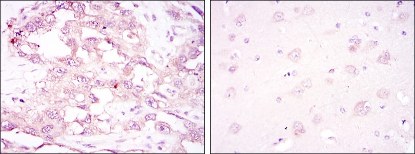
Figure 2: Immunohistochemical analysis of paraffin-embedded ovarian cancer (left) and brain tissues (right) using PRKAA1 mouse mAb with DAB staining.
Immunofluorescence analysis

Figure 3: Immunofluorescence analysis of NTERA-2 cells using PRKAA1 mouse mAb (green). Blue: DRAQ5 fluorescent DNA dye. Red: Actin filaments have been labeled with Alexa Fluor-555 phalloidin.
Flow cytometric

Figure 4: Flow cytometric analysis of PC-2 cells using PRKAA1 mouse mAb (green) and negative control (purple).
Elisa

Red: Control Antigen (100ng); Purple: Antigen (10ng); Green: Antigen (50ng); Blue: Antigen (100ng);
For Research Use Only. Not for use in diagnostic procedures.