

PRDM1 Primary Antibody
Item Information
Catalog #
Size
Price
Description
This gene encodes a protein that acts as a repressor of beta-interferon gene expression. The protein binds specifically to the PRDI (positive regulatory domain I element) of the beta-IFN gene promoter. Transcription of this gene increases upon virus induction. Two alternatively spliced transcript variants that encode different isoforms have been reported.
Product Overview
Entrez GenelD
639
Aliases
BLIMP1; PRDI-BF1
Clone#
2F1B6
Host / Isotype
Mouse / IgG2b
Species Reactivity
Human
Immunogen
Purified recombinant fragment of human PRDM1 (AA: 690-825) expressed in E. Coli.
Formulation
Purified antibody in PBS with 0.05% sodium azide
Storage
Store at 4°C short term. Aliquot and store at -20°C long term. Avoid freeze/thaw cycles.
Product Applications
WB (Western Blot)
1/500 - 1/2000
ICC (Immunocytochemistry)
1/200 - 1/1000
FCM (Flow Cytometry)
1/200 - 1/400
ELISA
1/10000
References
1.Proc Natl Acad Sci U S A. 2011 Dec 13;108(50):20119-24.
2.Mol Cancer Res. 2010 Jun;8(6):907-18.
2.Mol Cancer Res. 2010 Jun;8(6):907-18.
Product Image
Elisa

Figure 1: Black line: Control Antigen (100 ng); Purple line: Antigen(10ng); Blue line: Antigen (50 ng); Red line: Antigen (100 ng);
Western Blot
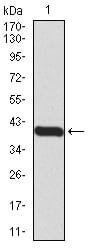
Figure 2:Western blot analysis using PRDM1 mAb against human PRDM1 (AA: 690-825) recombinant protein. (Expected MW is 40.3 kDa)
Western Blot

Figure 3:Western blot analysis using PRDM1 mAb against HEK293 (1) and PRDM1 (AA: 690-825)-hIgGFc transfected HEK293 (2) cell lysate.
Immunofluorescence analysis

Figure 4:Immunofluorescence analysis of HeLa cells using PRDM1 mouse mAb (green). Blue: DRAQ5 fluorescent DNA dye. Red: Actin filaments have been labeled with Alexa Fluor- 555 phalloidin. Secondary antibody from Fisher (Cat#: 35503)
Flow cytometric

Figure 5:Flow cytometric analysis of Raji cells using PRDM1 mouse mAb (green) and negative control (red).
For Research Use Only. Not for use in diagnostic procedures.