PPP2R4 Primary Antibody

Item Information
Catalog #Size/ConcentrationPrice
Specification
AliasesPP2A; PR53; PTPA
ProductOrderP
Clone#4D9
Entrez GenelD5524
FormulationPurified antibody in PBS with 0.05% sodium azide
HostMouse
IsotypeIgG1
ImmunogenPurified recombinant fragment of human PPP2R4 expressed in E. Coli.
MW41kDa
Shipping InformationThis product will ship in a box containing blue ice at a temperature of 4°C. Learn More
Species ReactivityHuman
Application
ELISA1/10000
ICC (Immunocytochemistry)1/200 - 1/1000
IHC_P(Immunohistochemistry)1/200 - 1/1000
FCM (Flow Cytometry)1/200 - 1/400
WB (Western Blot)1/500 - 1/2000
Sequence
1-154
Catalog#: 30308
Inquire
Contact us to order
Images
Western Blot
Figure 1: Western blot analysis using PPP2R4 mAb against human PPP2R4 (AA: 1-154) recombinant protein. (Expected MW is 41 kDa)
Immunohistochemical analysis
Figure 2: Immunohistochemical analysis of paraffin-embedded cervical cancer tissues using PPP2R4 mouse mAb with DAB staining.
Immunohistochemical analysis
Figure 3: Immunohistochemical analysis of paraffin-embedded lung cancer tissues using PPP2R4 mouse mAb with DAB staining.
Immunofluorescence analysis
Figure 4: Immunofluorescence analysis of Hela cells using PPP2R4 mouse mAb (green). Blue: DRAQ5 fluorescent DNA dye. Red: Actin filaments have been labeled with Alexa Fluor-555 phalloidin.
Flow cytometric
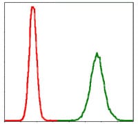
Figure 5: Flow cytometric analysis of MCF-7 cells using PPP2R4 mouse mAb (green) and negative control (red).
Elisa
Black line: Control Antigen (100 ng); Purple line: Antigen(10ng); Blue line: Antigen (50 ng); Red line: Antigen (100 ng);
Product Overview
Description

Protein phosphatase 2A is one of the four major Ser/Thr phosphatases and is implicated in the negative control of cell growth and division. Protein phosphatase 2A holoenzymes are heterotrimeric proteins composed of a structural subunit A, a catalytic subunit C, and a regulatory subunit B. The regulatory subunit is encoded by a diverse set of genes that have been grouped into the B/PR55, B'/PR61, and B''/PR72 families. These different regulatory subunits confer distinct enzymatic specificities and intracellular localizations to the holozenzyme. The product of this gene belongs to the B' family. This gene encodes a specific phosphotyrosyl phosphatase activator of the dimeric form of protein phosphatase 2A. Alternative splicing results in multiple transcript variants encoding different isoforms.

References (references)
References (references)1. Stem Cells. 2010 May;28(5):874-84.
2. Mol Biol Cell. 2010 Mar 15;21(6):1140-52.