
PPP1CB Primary Antibody
Item Information
Catalog #
Size
Price
Description
The protein encoded by this gene is one of the three catalytic subunits of protein phosphatase 1 (PP1). PP1 is a serine/threonine specific protein phosphatase known to be involved in the regulation of a variety of cellular processes, such as cell division, glycogen metabolism, muscle contractility, protein synthesis, and HIV-1 viral transcription. Mouse studies suggest that PP1 functions as a suppressor of learning and memory. Two alternatively spliced transcript variants encoding distinct isoforms have been observed
Product Overview
Entrez GenelD
5500
Aliases
PP1B; PP-1B; PPP1CD; PP1beta
Clone#
8A7C7
Host / Isotype
Mouse / IgG1
Species Reactivity
Human
Immunogen
Purified recombinant fragment of human PPP1CB (AA: 174-327) expressed in E. Coli.
Formulation
Purified antibody in PBS with 0.05% sodium azide.
Storage
Store at 4°C short term. Aliquot and store at -20°C long term. Avoid freeze/thaw cycles.
Product Applications
WB (Western Blot)
1/500 - 1/2000
IHC_P(Immunohistochemistry)
1/200 - 1/1000
ICC (Immunocytochemistry)
1/200 - 1/1000
FCM (Flow Cytometry)
1/200 - 1/400
ELISA
1/10000
References
1. J Biol Chem. 2011 Sep 23;286(38):32931-6.
2. Mol Biol Cell. 2010 Dec;21(24):4409-17.
2. Mol Biol Cell. 2010 Dec;21(24):4409-17.
Product Image
Western Blot

Figure 1: Western blot analysis using PPP1CB mAb against human PPP1CB (AA: 174-327) recombinant protein. (Expected MW is 43.2 kDa)
Western Blot
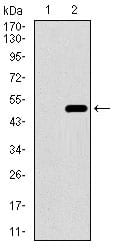
Figure 2: Western blot analysis using PPP1CB mAb against HEK293 (1) and PPP1CB (AA: 174-327)-hIgGFc transfected HEK293 (2) cell lysate.
Western Blot

Figure 3: Western blot analysis using PPP1CB mouse mAb against Jurkat (1), A431 (2), Hela (3), HepG2 (4), HEK293 (5), MCF-7 (6) cell lysate.
Immunofluorescence analysis
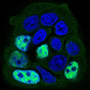
Figure 4: Immunofluorescence analysis of MCF-7 cells using PPP1CB mouse mAb (green). Blue: DRAQ5 fluorescent DNA dye. Secondary antibody from Fisher (Cat#: 35503)
Flow cytometric

Figure 5: Flow cytometric analysis of Jurkat cells using PPP1CB mouse mAb (green) and negative control (red).
Immunohistochemical analysis

Figure 6: Immunohistochemical analysis of paraffin-embedded cervical cancer tissues using PPP1CB mouse mAb with DAB staining.
Immunohistochemical analysis

Figure 7: Immunohistochemical analysis of paraffin-embedded rectum cancer tissues using PPP1CB mouse mAb with DAB staining.
Elisa
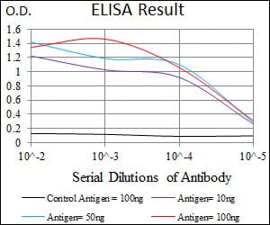
Black line: Control Antigen (100 ng); Purple line: Antigen(10ng); Blue line: Antigen (50 ng); Red line: Antigen (100 ng);
For Research Use Only. Not for use in diagnostic procedures.