
PPID Primary Antibody
Item Information
Catalog #
Size
Price
Description
The protein encoded by this gene is a member of the peptidyl-prolyl cis-trans isomerase (PPIase) family. PPIases catalyze the cis-trans isomerization of proline imidic peptide bonds in oligopeptides and accelerate the folding of proteins. This protein has been shown to possess PPIase activity and, similar to other family members, can bind to the immunosuppressant cyclosporin A.
Product Overview
Entrez GenelD
5481
Aliases
CYPD; CYP-40
Clone#
5D10E5
Host / Isotype
Mouse / Mouse IgG1
Species Reactivity
Human
Immunogen
Purified recombinant fragment of human PPID (AA: 171-370) expressed in E. Coli.
Formulation
Purified antibody in PBS with 0.05% sodium azide
Storage
Store at 4°C short term. Aliquot and store at -20°C long term. Avoid freeze/thaw cycles.
Product Applications
WB (Western Blot)
1/500 - 1/2000
IHC_P(Immunohistochemistry)
1/200 - 1/1000
FCM (Flow Cytometry)
1/200 - 1/400
ELISA
1/10000
References
1.Genet Mol Res. 2015 Apr 28;14(2):4258-68.
2.Oxid Med Cell Longev. 2019 Apr 4;2019:1729013.
2.Oxid Med Cell Longev. 2019 Apr 4;2019:1729013.
Product Image
Elisa
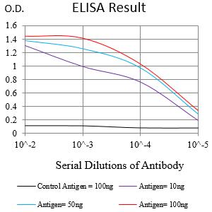
Figure 1:Black line: Control Antigen (100 ng);Purple line: Antigen (10ng); Blue line: Antigen (50 ng); Red line:Antigen (100 ng)
Western Blot

Figure 2:Western blot analysis using PPID mAb against human PPID (AA: 171-370) recombinant protein. (Expected MW is 25 kDa)
Western Blot
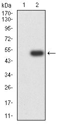
Figure 3:Western blot analysis using PPID mAb against HEK293-6e (1) and PPID (AA: 171-370)-hIgGFc transfected HEK293-6e (2) cell lysate.
Immunofluorescence analysis

Figure 4:Flow cytometric analysis of Jurkat cells using PPID mouse mAb (green) and negative control (red).
Immunohistochemical analysis

Figure 5:Immunohistochemical analysis of paraffin-embedded bladder cancer tissues using PPID mouse mAb with DAB staining.
Immunohistochemical analysis

Figure 6:Immunohistochemical analysis of paraffin-embedded prostate cancer tissues using PPID mouse mAb with DAB staining.
Immunohistochemical analysis

Figure 7:Immunohistochemical analysis of paraffin-embedded breast cancer tissues using PPID mouse mAb with DAB staining.
For Research Use Only. Not for use in diagnostic procedures.