

POU5F1 Primary Antibody
Item Information
Catalog #
Size
Price
Description
This gene encodes a transcription factor containing a POU homeodomain that plays a key role in embryonic development and stem cell pluripotency. Aberrant expression of this gene in adult tissues is associated with tumorigenesis. This gene can participate in a translocation with the Ewing's sarcoma gene on chromosome 21, which also leads to tumor formation. Alternative splicing, as well as usage of alternative AUG and non-AUG translation initiation codons, results in multiple isoforms. One of the AUG start codons is polymorphic in human populations. Related pseudogenes have been identified on chromosomes 1, 3, 8, 10, and 12.
Product Overview
Entrez GenelD
5460
Aliases
OCT3; OCT4; OTF3; OTF4; OTF-3; Oct-3; Oct-4
Clone#
3C1F5
Host / Isotype
Mouse / Mouse IgG1
Species Reactivity
Human
Immunogen
Purified recombinant fragment of human (AA: 136-360) expressed in E. Coli.
Formulation
Purified antibody in PBS with 0.05% sodium azide
Storage
Store at 4°C short term. Aliquot and store at -20°C long term. Avoid freeze/thaw cycles.
Product Applications
WB (Western Blot)
1/500 - 1/2000
IHC_P(Immunohistochemistry)
1/200 - 1/1000
FCM (Flow Cytometry)
1/200 - 1/400
ELISA
1/10000
References
1.Biomark Med. 2020 Oct;14(15):1473-1484.
2.Cancer Med. 2020 Dec;9(23):8782-8800.
2.Cancer Med. 2020 Dec;9(23):8782-8800.
Product Image
Elisa

Figure 1:Black line: Control Antigen (100 ng);Purple line: Antigen (10ng); Blue line: Antigen (50 ng); Red line:Antigen (100 ng)
Western Blot
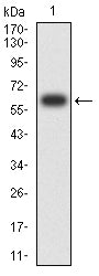
Figure 2:Western blot analysis using POU5F1 mAb against human POU5F1 (AA: 136-360) recombinant protein. (Expected MW is 65.2 kDa)
Western Blot

Figure 3:Western blot analysis using POU5F1 mAb against HEK293-6e (1) and POU5F1 (AA: 136-360)-hIgGFc transfected HEK293-6e (2) cell lysate.
Immunofluorescence analysis

Figure 4:Flow cytometric analysis of Hela cells using POU5F1 mouse mAb (green) and negative control (red).
Immunofluorescence analysis

Figure 5:Flow cytometric analysis of K562 cells using POU5F1 mouse mAb (green) and negative control (red).
Immunohistochemical analysis
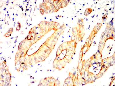
Figure 6:Immunohistochemical analysis of paraffin-embedded colon cancer tissues using POU5F1 mouse mAb with DAB staining.
Immunohistochemical analysis

Figure 7:Immunohistochemical analysis of paraffin-embedded rectum cancer tissues using POU5F1 mouse mAb with DAB staining.
For Research Use Only. Not for use in diagnostic procedures.