

PON1 Primary Antibody
Item Information
Catalog #
Size
Price
Description
The enzyme encoded by this gene is an arylesterase that mainly hydrolyzes paroxon to produce p-nitrophenol. Paroxon is an organophosphorus anticholinesterase compound that is produced in vivo by oxidation of the insecticide parathion. Polymorphisms in this gene are a risk factor in coronary artery disease. The gene is found in a cluster of three related paraoxonase genes at 7q21.3.
Product Overview
Entrez GenelD
5444
Aliases
ESA; PON; MVCD5
Clone#
4G8A12
Host / Isotype
Mouse / IgG1
Species Reactivity
Human
Immunogen
Purified recombinant fragment of human PON1 (AA: 20-155) expressed in E. Coli.
Formulation
Ascitic fluid containing 0.03% sodium azide.
Storage
Store at 4°C short term. Aliquot and store at -20°C long term. Avoid freeze/thaw cycles.
Product Applications
WB (Western Blot)
1/500 - 1/2000
IHC_P(Immunohistochemistry)
1/200 - 1/1000
FCM (Flow Cytometry)
1/200 - 1/400
ELISA
1/10000
References
1. Redox Rep. 2012;17(5):214-8.
2. Cancer Epidemiol. 2012 Apr;36(2):e101-3.
2. Cancer Epidemiol. 2012 Apr;36(2):e101-3.
Product Image
Western Blot

Figure 1: Western blot analysis using PON1 mAb against human PON1 recombinant protein. (Expected MW is 40.6 kDa)
Western Blot

Figure 2: Western blot analysis using PON1 mAb against HEK293 (1) and PON1 (AA: 20-155)-hIgGFc transfected HEK293 (2) cell lysate.
Western Blot

Figure 3: Western blot analysis using PON1 mouse mAb against human plasma cell lysate.
Flow cytometric

Figure 4: Flow cytometric analysis of Hela cells using PON1 mouse mAb (green) and negative control (red).
Immunohistochemical analysis

Figure 5: Immunohistochemical analysis of paraffin-embedded rectum cancer tissues using PON1 mouse mAb with DAB staining.
Immunohistochemical analysis

Figure 6: Immunohistochemical analysis of paraffin-embedded liver cancer tissues using PON1 mouse mAb with DAB staining.
Elisa
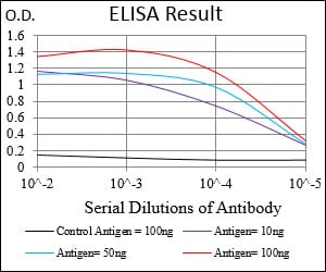
Black line: Control Antigen (100 ng); Purple line: Antigen(10ng); Blue line: Antigen (50 ng); Red line: Antigen (100 ng);
For Research Use Only. Not for use in diagnostic procedures.