
Mouse Monoclonal Antibody to PMEL
Item Information
Catalog #
Size
Price
Description
This gene encodes a melanocyte-specific type I transmembrane glycoprotein. The encoded protein is enriched in melanosomes, which are the melanin-producing organelles in melanocytes, and plays an essential role in the structural organization of premelanosomes. This protein is involved in generating internal matrix fibers that define the transition from Stage I to Stage II melanosomes. This protein undergoes a complex pattern of prosttranslational processing and modification that is essential to the proper functioning of the protein. A secreted form of this protein that is released by proteolytic ectodomain shedding may be used as a melanoma-specific serum marker. Alternate splicing results in multiple transcript variants.
Product Overview
Entrez GenelD
6490
Aliases
P1; SI; SIL; ME20; P100; SILV; ME20M; gp100; ME20-M; PMEL17; D12S53E
Clone#
1D4A5
Host / Isotype
Mouse / IgG1
Immunogen
Purified recombinant fragment of human PMEL (AA: 25-192) expressed in E. Coli.
Formulation
Purified antibody in PBS with 0.05% sodium azide
Storage
Store at 4°C short term. Aliquot and store at -20°C long term. Avoid freeze/thaw cycles.
Product Applications
WB (Western Blot)
1/500 - 1/2000
FCM (Flow Cytometry)
1/200 - 1/400
ELISA
1/10000
References
1.J Biol Chem. 2020 May 22;295(21):7544-7553. 2.Biochem Biophys Res Commun. 2018 Sep 18;503(4):2536-2542.
Product Image
Elisa
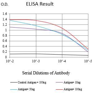
Figure 1:Black line: Control Antigen (100 ng);Purple line: Antigen (10ng); Blue line: Antigen (50 ng); Red line:Antigen (100 ng)
Western Blot

Figure 2:Western blot analysis using PMEL mAb against human PMEL (AA: 25-192) recombinant protein. (Expected MW is 44.7 kDa)
Western Blot

Figure 3:Western blot analysis using PMEL mAb against HEK293-6e (1) and PMEL (AA: 25-192)-hIgGFc transfected HEK293-6e (2) cell lysate.
Flow cytometric analysis

Figure 4:Flow cytometric analysis of B16 cells using PMEL mouse mAb (green) and negative control (red).
For Research Use Only. Not for use in diagnostic procedures.