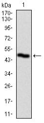
PLK1 Primary Antibody

Item Information
Catalog #
Size
Price
Description

PLK1 is critical for the initiation of centrosome maturation. Polo-like kinases (PLKs) are a family of four serine/threonine protein kinases that are critical regulators of cell cycle progression, mitosis, cytokinesis, and the DNA damage response. PLK1, -2 and -3 are ubiquitously expressed, whereas PLK4 is restricted to a few tissues including the testes and the thymus. The mRNA and protein expression of PLK1, -2 and -4 are coordinately regulated during cell cycle progression, but PLK3 levels are independent of the other three family members. Furthermore, PLK3 is a much more stable protein than PLK1, -2 or -4. PLK1 is the most well characterized member of this family and strongly promotes the progression of cells through mitosis. During the various stages of mitosis PLK1 localizes to the centrosomes, kinetochores and central spindle. PLKs are dysregulated in a variety of human cancers. PLK1 overexpression correlates with cellular proliferation and poor prognosis. PLK2 and PLK3 are involved in checkpoint-mediated cell cycle arrest to ensure genetic stability. Loss-of-function mutations in these enzymes can lead to oncogenic transformation.

Product Overview
Entrez GenelD
5347
Aliases
PLK; STPK13
Clone#
1D1
Host / Isotype
Mouse / IgG1
Species Reactivity
Human
Immunogen
Purified recombinant fragment of human PLK1 (AA: 331-508) expressed in E. Coli.
Formulation
Purified antibody in PBS with 0.05% sodium azide
Storage
4°C; -20°C for long term storage
Product Applications
WB (Western Blot)
1/500 - 1/2000
ICC (Immunocytochemistry)
1/50
ELISA
1/10000
References
1.J Cell Biol. 2011 Dec 26;195(7):1093-101.
2.Eur J Cancer. 2011 Sep;47(14):2166-74.
Product Image
Western Blot
Figure 1: Western blot analysis using PLK1 mAb against human PLK1 recombinant protein. (Expected MW is 45.7 kDa)
Immunofluorescence analysis
Figure 2: Immunofluorescence analysis of HeLa cells using PLK1 mouse mAb (green). Blue: DRAQ5 fluorescent DNA dye. Red: Actin filaments have been labeled with Alexa Fluor-555 phalloidin.
Elisa
Black line: Control Antigen (100 ng); Purple line: Antigen(10ng); Blue line: Antigen (50 ng); Red line: Antigen (100 ng);
For Research Use Only. Not for use in diagnostic procedures.