
PLIN3 Primary Antibody
Item Information
Catalog #
Size
Price
Description
Mannose 6-phophate receptors (MPRs) deliver lysosomal hydrolase from the Golgi to endosomes and then return to the Golgi complex. The protein encoded by this gene interacts with the cytoplasmic domains of both cation-independent and cation-dependent MPRs, and is required for endosome-to-Golgi transport. This protein also binds directly to the GTPase RAB9 (RAB9A), a member of the RAS oncogene family. The interaction with RAB9 has been shown to increase the affinity of this protein for its cargo. Multiple transcript variants encoding different isoforms have been found for this gene.
Product Overview
Entrez GenelD
10226
Aliases
PP17; TIP47; M6PRBP1
Clone#
6C9D9
Host / Isotype
Mouse / Mouse IgG2a
Species Reactivity
Human
Immunogen
Purified recombinant fragment of human PLIN3 expressed in E. Coli.
Formulation
Purified antibody in PBS with 0.05% sodium azide
Storage
Store at 4°C short term. Aliquot and store at -20°C long term. Avoid freeze/thaw cycles.
Product Applications
WB (Western Blot)
1/500 - 1/2000
ICC (Immunocytochemistry)
1/100 - 1/500
FCM (Flow Cytometry)
1/200 - 1/400
References
1.Med Oncol. 2021 Aug 19;38(10):116.
2.Urol Oncol. 2018 Jul;36(7):343.e9-343.e19.
2.Urol Oncol. 2018 Jul;36(7):343.e9-343.e19.
Product Image
Western Blot
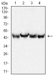
Figure 1:Western blot analysis using PLIN3 mouse mAb against THP-1 (1), HepG2 (2), K562 (3) and Hela (4) cell lysate.
Immunohistochemical analysis

Figure 2:Immunofluorescence analysis of Hela cells using PLIN3 mouse mAb (green). Blue: DRAQ5 fluorescent DNA dye. Red: Actin filaments have been labeled with Alexa Fluor- 555 phalloidin. Secondary antibody from Fisher (Cat#: 35503)
Immunofluorescence analysis

Figure 3:Flow cytometric analysis of Hela cells using PLIN3 mouse mAb (green) and negative control (red).
For Research Use Only. Not for use in diagnostic procedures.