

PLD2 Primary Antibody
Item Information
Catalog #
Size
Price
Description
The protein encoded by this gene catalyzes the hydrolysis of phosphatidylcholine to phosphatidic acid and choline. The activity of the encoded enzyme is enhanced by phosphatidylinositol 4,5-bisphosphate and ADP-ribosylation factor-1. This protein localizes to the peripheral membrane and may be involved in cytoskeletal organization, cell cycle control, transcriptional regulation, and/or regulated secretion. Two transcript variants encoding different isoforms have been found for this gene.
Product Overview
Entrez GenelD
5338
Aliases
N
Clone#
7E4D9
Host / Isotype
Mouse / IgG1
Species Reactivity
Human
Immunogen
Purified recombinant fragment of human PLD2 (AA: 834-933) expressed in E. Coli.
Formulation
Purified antibody in PBS with 0.05% sodium azide
Storage
Store at 4°C short term. Aliquot and store at -20°C long term. Avoid freeze/thaw cycles.
Product Applications
WB (Western Blot)
1/500 - 1/2000
IHC_P(Immunohistochemistry)
1/200 - 1/1000
ICC (Immunocytochemistry)
1/200 - 1/1000
FCM (Flow Cytometry)
1/200 - 1/400
ELISA
1/10000
References
1.Exp Mol Med. 2014 Dec 5;46:e124.
2.FEBS Lett. 2014 Aug 25;588(17):3251-8.
2.FEBS Lett. 2014 Aug 25;588(17):3251-8.
Product Image
Elisa
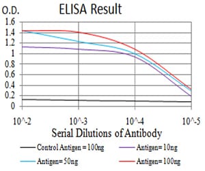
Figure 1: Black line: Control Antigen (100 ng);Purple line: Antigen (10ng); Blue line: Antigen (50 ng); Red line:Antigen (100 ng)
Western Blot
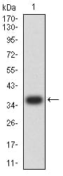
Figure 2:Western blot analysis using PLD2 mAb against human PLD2 (AA: 834-933) recombinant protein. (Expected MW is 37.4 kDa)
Western Blot
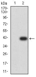
Figure 3:Western blot analysis using PLD2 mAb against HEK293 (1) and PLD2 (AA: 834-933)-hIgGFc transfected HEK293 (2) cell lysate.
Immunofluorescence analysis
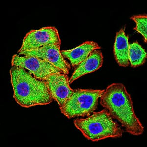
Figure 4:Immunofluorescence analysis of MCF-7 cells using PLD2 mouse mAb (green). Blue: DRAQ5 fluorescent DNA dye. Red: Actin filaments have been labeled with Alexa Fluor- 555 phalloidin. Secondary antibody from Fisher (Cat#: 35503)
Immunofluorescence analysis
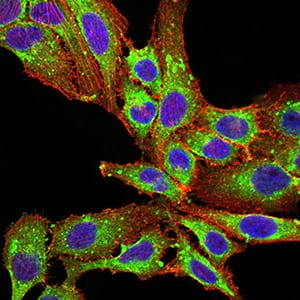
Figure 5:Immunofluorescence analysis of SK-OV-3 cells using PLD2 mouse mAb (green). Blue: DRAQ5 fluorescent DNA dye. Red: Actin filaments have been labeled with Alexa Fluor- 555 phalloidin. Secondary antibody from Fisher (Cat#: 35503)
Flow cytometric
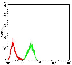
Figure 6:Flow cytometric analysis of Hela cells using PLD2 mouse mAb (green) and negative control (red).
Immunohistochemical analysis
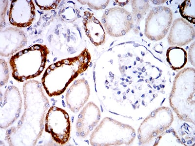
Figure 7:Immunohistochemical analysis of paraffin-embedded renal tissues using PLD2 mouse mAb with DAB staining.
Immunohistochemical analysis
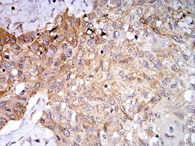
Figure 8:Immunohistochemical analysis of paraffin-embedded esophageal cancer tissues using PLD2 mouse mAb with DAB staining.
For Research Use Only. Not for use in diagnostic procedures.