

PLCG2 Primary Antibody
Item Information
Catalog #
Size
Price
Description
The protein encoded by this gene is a transmembrane signaling enzyme that catalyzes the conversion of 1-phosphatidyl-1D-myo-inositol 4,5-bisphosphate to 1D-myo-inositol 1,4,5-trisphosphate (IP3) and diacylglycerol (DAG) using calcium as a cofactor. IP3 and DAG are second messenger molecules important for transmitting signals from growth factor receptors and immune system receptors across the cell membrane. Mutations in this gene have been found in autoinflammation, antibody deficiency, and immune dysregulation syndrome and familial cold autoinflammatory syndrome 3.
Product Overview
Entrez GenelD
5336
Aliases
FCAS3; APLAID; PLC-IV; PLC-gamma-2
Clone#
1E10C11
Host / Isotype
Mouse / IgG1
Species Reactivity
Human
Immunogen
Purified recombinant fragment of human PLCG2 (AA: 826-985) expressed in E. Coli.
Formulation
Purified antibody in PBS with 0.05% sodium azide
Storage
Store at 4°C short term. Aliquot and store at -20°C long term. Avoid freeze/thaw cycles.
Product Applications
WB (Western Blot)
1/500 - 1/2000
IHC_P(Immunohistochemistry)
1/200 - 1/1000
ELISA
1/10000
References
1.J Biol Chem. 2005 Nov 25;280(47):38923-31.
2.World J Gastroenterol. 2003 Nov;9(11):2413-8.
2.World J Gastroenterol. 2003 Nov;9(11):2413-8.
Product Image
Elisa
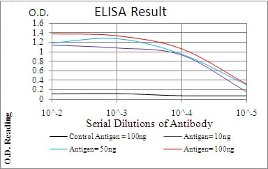
Figure 1: Black line: Control Antigen (100 ng); Purple line: Antigen(10ng); Blue line: Antigen (50 ng); Red line: Antigen (100 ng);
Western Blot
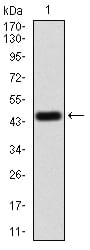
Figure 2:Western blot analysis using PLCG2 mAb against human PLCG2 (AA: 826-985) recombinant protein. (Expected MW is 44.6 kDa)
Western Blot
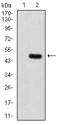
Figure 3:Western blot analysis using PLCG2 mAb against HEK293 (1) and PLCG2 (AA: 826-985)-hIgGFc transfected HEK293 (2) cell lysate.
Immunohistochemical analysis

Figure 4:Immunohistochemical analysis of paraffin-embedded Hela tissues using PLCG2 mouse mAb with DAB staining.
For Research Use Only. Not for use in diagnostic procedures.