
PINCH Primary Antibody
Item Information
Catalog #
Size
Price
Description
The protein encoded by this gene is an adaptor protein which contains five LIM domains, or double zinc fingers. The protein is likely involved in integrin signaling through its LIM domain-mediated interaction with integrin-linked kinase, found in focal adhesion plaques. It is also thought to act as a bridge linking integrin-linked kinase to NCK adaptor protein 2, which is involved in growth factor receptor kinase signaling pathways. Its localization to the periphery of spreading cells also suggests that this protein may play a role in integrin-mediated cell adhesion or spreading. Several transcript variants encoding different isoforms have been found for this gene.
Product Overview
Entrez GenelD
3987
Aliases
PINCH; PINCH1; PINCH-1; LIMS1
Clone#
5G7
Host / Isotype
Mouse / IgG1
Species Reactivity
Human
Immunogen
Purified recombinant fragment of human PINCH expressed in E. Coli.
Formulation
Ascitic fluid containing 0.03% sodium azide.
Storage
Store at 4°C short term. Aliquot and store at -20°C long term. Avoid freeze/thaw cycles.
Product Applications
WB (Western Blot)
1/500 - 1/2000
ICC (Immunocytochemistry)
1/200 - 1/1000
FCM (Flow Cytometry)
1/200 - 1/400
ELISA
1/10000
References
1. J Biol Chem. 2009 Feb 27;284(9):5836-44.
2. Proc Natl Acad Sci U S A. 2008 Dec 30;105(52):20677-82.
2. Proc Natl Acad Sci U S A. 2008 Dec 30;105(52):20677-82.
Product Image
Western Blot

Figure 1: Western blot analysis using PINCH mAb against human PINCH (AA: 87-249) recombinant protein. (Expected MW is 44.2 kDa)
Western Blot
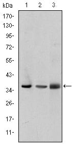
Figure 2: Western blot analysis using PINCH mouse mAb against A549 (1), Jurkat (2), and Hela (3) cell lysate.
Immunofluorescence analysis

Figure 3: Immunofluorescence analysis of HepG2 cells using PINCH mouse mAb (green). Blue: DRAQ5 fluorescent DNA dye. Red: Actin filaments have been labeled with Alexa Fluor-555 phalloidin.
Flow cytometric

Figure 4: Flow cytometric analysis of Hela cells using PINCH mouse mAb (blue) and negative control (red).
Elisa

Red: Control Antigen (100ng); Purple: Antigen (10ng); Green: Antigen (50ng); Blue: Antigen (100ng);
For Research Use Only. Not for use in diagnostic procedures.