
PIK3CA Primary Antibody
Item Information
Catalog #
Size
Price
Description
Phosphatidylinositol 3-kinase is composed of an 85 kDa regulatory subunit and a 110 kDa catalytic subunit. The protein encoded by this gene represents the catalytic subunit, which uses ATP to phosphorylate PtdIns, PtdIns4P and PtdIns(4,5)P2. This gene has been found to be oncogenic and has been implicated in cervical cancers.
Product Overview
Entrez GenelD
5290
Aliases
PI3K; p110-alpha
Clone#
4F3
Host / Isotype
Mouse / IgG1
Species Reactivity
Human
Immunogen
Purified recombinant fragment of human PIK3CA expressed in E. Coli.
Formulation
Purified antibody in PBS with 0.05% sodium azide
Storage
4°C; -20°C for long term storage
Product Applications
WB (Western Blot)
1/500 - 1/2000
ICC (Immunocytochemistry)
1/200 - 1/1000
FCM (Flow Cytometry)
1/200 - 1/400
ELISA
1/10000
References
Cancer Res. 2009 Dec 1;69(23):8868-76.
Diagn Mol Pathol. 2009 Dec;18(4):200-5.
Diagn Mol Pathol. 2009 Dec;18(4):200-5.
Product Image
Western Blot

Figure 1: Western blot analysis using PIK3CA mAb against human PIK3CA (AA: 881-1068) recombinant protein. (Expected MW is 47.4 kDa)
Western Blot
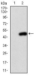
Figure 2: Western blot analysis using PIK3CA mAb against HEK293 (1) and PIK3CA (AA: 881-1068)-hIgGFc transfected HEK293 (2) cell lysate.
Immunofluorescence analysis

Figure 3: Immunofluorescence analysis of HeLa cells using PIK3CA mouse mAb (green). Blue: DRAQ5 fluorescent DNA dye.
Flow cytometric

Figure 4: Flow cytometric analysis of Jurkat cells using PIK3CA mouse mAb (green) and negative control (red).
Elisa
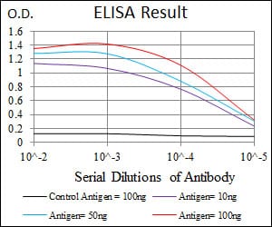
Black line: Control Antigen (100 ng); Purple line: Antigen(10ng); Blue line: Antigen (50 ng); Red line: Antigen (100 ng);
For Research Use Only. Not for use in diagnostic procedures.