

PIGR Primary Antibody
Item Information
Catalog #
Size
Price
Description
This gene is a member of the immunoglobulin superfamily. The encoded poly-Ig receptor binds polymeric immunoglobulin molecules at the basolateral surface of epithelial cells; the complex is then transported across the cell to be secreted at the apical surface. A significant association was found between immunoglobulin A nephropathy and several SNPs in this gene.
Product Overview
Entrez GenelD
5284
Clone#
6E5A9
Host / Isotype
Mouse / IgG1
Species Reactivity
Human
Immunogen
Purified recombinant fragment of human PIGR (AA: extra 19-170) expressed in E. Coli.
Formulation
Purified antibody in PBS with 0.05% sodium azide
Storage
Store at 4°C short term. Aliquot and store at -20°C long term. Avoid freeze/thaw cycles.
Product Applications
WB (Western Blot)
1/500 - 1/2000
IHC_P(Immunohistochemistry)
1/200 - 1/1000
ELISA
1/10000
References
1.Pancreatology. 2017 Mar - Apr;17(2):295-302.2.PLoS One. 2014 Nov 14;9(11):e112728.
Product Image
Elisa
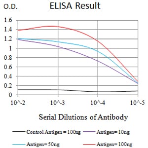
Figure 1: Black line: Control Antigen (100 ng);Purple line: Antigen (10ng); Blue line: Antigen (50 ng); Red line:Antigen (100 ng)
Western Blot
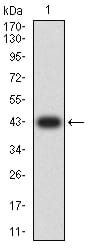
Figure 2:Western blot analysis using PIGR mAb against human PIGR (AA: extra 19-170) recombinant protein. (Expected MW is 42.6 kDa)
Western Blot
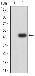
Figure 3:Western blot analysis using PIGR mAb against HEK293 (1) and PIGR (AA: extra 19-170)-hIgGFc transfected HEK293 (2) cell lysate.
Immunohistochemical analysis
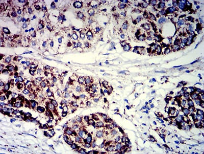
Figure 4:Immunohistochemical analysis of paraffin-embedded ovarian cancer tissues using PIGR mouse mAb with DAB staining.
Immunohistochemical analysis
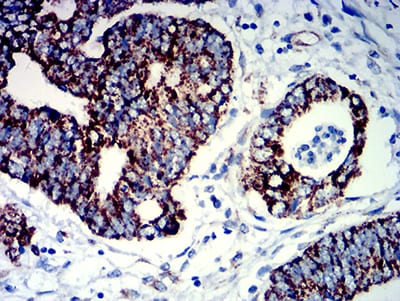
Figure 5:Immunohistochemical analysis of paraffin-embedded rectum cancer tissues using PIGR mouse mAb with DAB staining.
For Research Use Only. Not for use in diagnostic procedures.