

Phospho-4E-BP1 (Ser65) Primary Antibody
Item Information
Catalog #
Size
Price
Description
This gene encodes one member of a family of translation repressor proteins. The protein directly interacts with eukaryotic translation initiation factor 4E (eIF4E), which is a limiting component of the multisubunit complex that recruits 40S ribosomal subunits to the 5' end of mRNAs. Interaction of this protein with eIF4E inhibits complex assembly and represses translation. This protein is phosphorylated in response to various signals including UV irradiation and insulin signaling, resulting in its dissociation from eIF4E and activation of mRNA translation.
Product Overview
Entrez GenelD
1978
Aliases
EIF4EBP1; BP-1; 4EBP1; 4E-BP1; PHAS-I
Clone#
2D1G11
Host / Isotype
Mouse / IgG1
Species Reactivity
Human
Immunogen
Synthesized peptide of human Phospho-4E-BP1 (Ser65).
Formulation
Purified antibody in PBS with 0.05% sodium azide
Storage
Store at 4°C short term. Aliquot and store at -20°C long term. Avoid freeze/thaw cycles.
Product Applications
IHC_P(Immunohistochemistry)
1/200 - 1/1000
FCM (Flow Cytometry)
1/200 - 1/400
ELISA
1/10000
References
1.Sci Signal. 2015 Nov 17;8(403):ra116.
2.Oncotarget. 2015 Sep 15;6(27):24092-104.
2.Oncotarget. 2015 Sep 15;6(27):24092-104.
Product Image
Elisa

Figure 1: Black line: Control Antigen (100 ng);Purple line: Antigen (10ng); Blue line: Antigen (50 ng); Red line:Antigen (100 ng)
Flow cytometric

Figure 2:Flow cytometric analysis of Jurkat cells using Phospho-4E-BP1 (Ser65) mouse mAb (green) and negative control (red).
Immunohistochemical analysis
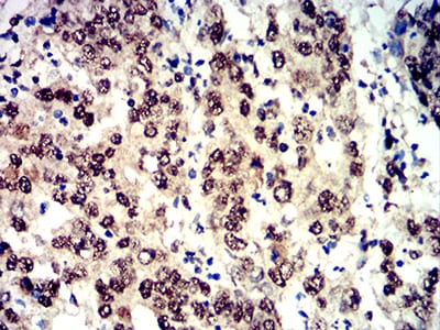
Figure 3:Immunohistochemical analysis of paraffin-embedded stomach cancer tissues using Phospho-4E-BP1 (Ser65) mouse mAb with DAB staining.
Immunohistochemical analysis

Figure 4:Immunohistochemical analysis of paraffin-embedded rectum cancer tissues using Phospho-4E-BP1 (Ser65) mouse mAb with DAB staining.
For Research Use Only. Not for use in diagnostic procedures.