

PHC1 Primary Antibody
Item Information
Catalog #
Size
Price
Description
This gene is a homolog of the Drosophila polyhomeotic gene, which is a member of the Polycomb group of genes. The gene product is a component of a multimeric protein complex that contains EDR2 and the vertebrate Polycomb protein BMH1. The gene product, the EDR2 protein, and the Drosophila polyhomeotic protein share 2 highly conserved domains, named homology domains I and II. These domains are involved in protein-protein interactions and may mediate heterodimerization of the protein encoded by this gene and the EDR2 protein.
Product Overview
Entrez GenelD
1911
Aliases
EDR1; HPH1; RAE28
Clone#
1F3F3
Host / Isotype
Mouse / IgG1
Species Reactivity
Human
Immunogen
Purified recombinant fragment of human PHC1 (AA: 758-1004) expressed in E. Coli.
Formulation
Purified antibody in PBS with 0.05% sodium azide
Storage
Store at 4°C short term. Aliquot and store at -20°C long term. Avoid freeze/thaw cycles.
Product Applications
WB (Western Blot)
1/500 - 1/2000
FCM (Flow Cytometry)
1/200 - 1/400
ELISA
1/10000
References
1.Development. 2007 Feb;134(3):579-90.
2.Nature. 2006 May 18;441(7091):349-53.
2.Nature. 2006 May 18;441(7091):349-53.
Product Image
Western Blot

Figure 1: Western blot analysis using PHC1 mAb against human PHC1 recombinant protein. (Expected MW is 52.8 kDa)
Western Blot
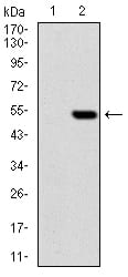
Figure 2: Western blot analysis using PHC1 mAb against HEK293 (1) and PHC1 (AA: 758-1004)-hIgGFc transfected HEK293 (2) cell lysate.
Flow cytometric

Figure 3: Flow cytometric analysis of HEK293 cells using PHC1 mouse mAb (green) and negative control (red).
Elisa

Black line: Control Antigen (100 ng); Purple line: Antigen(10ng); Blue line: Antigen (50 ng); Red line: Antigen (100 ng);
For Research Use Only. Not for use in diagnostic procedures.