
PGR Primary Antibody
Item Information
Catalog #
Size
Price
Description
This gene encodes a member of the steroid receptor superfamily. The encoded protein mediates the physiological effects of progesterone, which plays a central role in reproductive events associated with the establishment and maintenance of pregnancy. This gene uses two distinct promotors and translation start sites in the first exon to produce several transcript variants, both protein coding and non-protein coding. Two of the isoforms (A and B) are identical except for an additional 165 amino acids found in the N-terminus of isoform B and mediate their own response genes and physiologic effects with little overlap. [provided by RefSeq, Sep 2015]
Product Overview
Entrez GenelD
5241
Aliases
PR; NR3C3
Clone#
2E9F2
Host / Isotype
Mouse / Mouse IgG1
Immunogen
Purified recombinant fragment of human PGR (AA: 166-411) expressed in E. Coli.
Formulation
Purified antibody in PBS with 0.05% sodium azide
Storage
Store at 4°C short term. Aliquot and store at -20°C long term. Avoid freeze/thaw cycles.
Product Applications
WB (Western Blot)
1/500 - 1/2000
IHC_P(Immunohistochemistry)
1/200-1/1000
FCM (Flow Cytometry)
1/200-1/400
ELISA
1/10000
References
1,Am J Surg Pathol. 2019 Jun;43(6):810-818. 2,J Steroid Biochem Mol Biol. 2019 Jun;190:212-223.
Product Image
ELISA

Figure 1: Black line: Control Antigen (100 ng);Purple line: Antigen (10ng); Blue line: Antigen (50 ng); Red line: Antigen (100 ng)
WESTERN BLOT

Figure 2: Western blot analysis using PGR mAb against human PGR (AA: 166-411) recombinant protein. (Expected MW is 27.8 kDa)
WESTERN BLOT
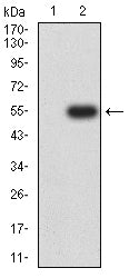
Figure 3: Western blot analysis using PGR mAb against HEK293-6e (1) and PGR (AA: 166-411)-hIgGFc transfected HEK293-6e (2) cell lysate.
WESTERN BLOT
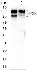
Figure 4: Western blot analysis using PGR mouse mAb against T47D (1), and C2C12 (2) cell lysate.
FLOW CYTOMETRY
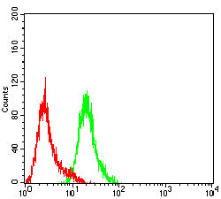
Figure 5: Flow cytometric analysis of Hela cells using PGR mouse mAb (green) and negative control (red).
IMMUNOHISTOCHEMISTRY
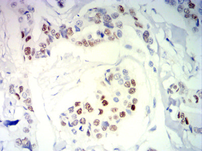
Figure 6: Immunohistochemical analysis of paraffin-embedded breast cancer tissues using PGR mouse mAb with DAB staining.
IMMUNOHISTOCHEMISTRY

Figure 7: Immunohistochemical analysis of paraffin-embedded cervical cancer tissues using PGR mouse mAb with DAB staining.
For Research Use Only. Not for use in diagnostic procedures.