

PDXK Primary Antibody
Item Information
Catalog #
Size
Price
Description
The protein encoded by this gene phosphorylates vitamin B6, a step required for the conversion of vitamin B6 to pyridoxal-5-phosphate, an important cofactor in intermediary metabolism. The encoded protein is cytoplasmic and probably acts as a homodimer. Alternatively spliced transcript variants have been described, but their biological validity has not been determined.
Product Overview
Entrez GenelD
8566
Aliases
PKH; PNK; HMSN6C; PRED79; C21orf97; HEL-S-1a; C21orf124
Clone#
4B4G3
Host / Isotype
Mouse / Mouse IgG1
Species Reactivity
Human, Mouse
Immunogen
Purified recombinant fragment of human PDXK (AA:1-312) expressed in E. Coli.
Formulation
Purified antibody in PBS with 0.05% sodium azide
Storage
Store at 4°C short term. Aliquot and store at -20°C long term. Avoid freeze/thaw cycles.
Product Applications
WB (Western Blot)
1/500 - 1/2000
IHC_P(Immunohistochemistry)
1/200 - 1/1000
ICC (Immunocytochemistry)
1/50 - 1/200
FCM (Flow Cytometry)
1/200 - 1/400
References
1.Ann Neurol. 2010 Mar;67(3):411-2; author reply 412.
2.Xi Bao Yu Fen Zi Mian Yi Xue Za Zhi. 2020 Jun;36(6):542-548.
2.Xi Bao Yu Fen Zi Mian Yi Xue Za Zhi. 2020 Jun;36(6):542-548.
Product Image
Western Blot

Figure 1:Western blot analysis using PDXK mouse mAb against Hela (1), HepG2 (2), MCF-7 (3), HEK293 (4), mouse liver (5) and mouse kidney (6) cell lysate.
Immunohistochemical analysis

Figure 2:Immunofluorescence analysis of Hala cells using PDXK mouse mAb (green). Blue: DRAQ5 fluorescent DNA dye. Red: Actin filaments have been labeled with Alexa Fluor- 555 phalloidin. Secondary antibody from Fisher (Cat#: 35503)
Immunofluorescence analysis
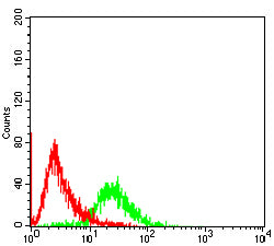
Figure 3:Flow cytometric analysis of HEK293 cells using PDXK mouse mAb (green) and negative control (red).
Immunohistochemical analysis

Figure 4:Immunohistochemical analysis of paraffin-embedded cervical cancer tissues using PDXK mouse mAb with DAB staining.
Immunohistochemical analysis

Figure 5:Immunohistochemical analysis of paraffin-embedded colon cancer tissues using PDXK mouse mAb with DAB staining.
For Research Use Only. Not for use in diagnostic procedures.