
PDPK1 Primary Antibody
Item Information
Catalog #
Size
Price
Description
Phosphoinositide-dependent kinase 1 (PDPK1, PDK1) is a serine/threonine protein kinase integral to the function of the PI 3-K/Akt signaling pathway. PDK1 and mTORC2 both phosphorylate and activate PKB/Akt, ensuring a cellular response to stimuli such as growth factors and insulin signaling. Akt is the main effector of PDK1.
Product Overview
Entrez GenelD
5170
Aliases
PDK1; PDPK2; PDPK2P; PRO0461
Clone#
3H3D9
Host / Isotype
Mouse / IgG1
Species Reactivity
Human
Immunogen
Purified recombinant fragment of human PDPK1 (AA: 457-556 expressed in E. Coli.
Formulation
Purified antibody in PBS with 0.05% sodium azide
Storage
Store at 4°C short term. Aliquot and store at -20°C long term. Avoid freeze/thaw cycles.
Product Applications
WB (Western Blot)
1/500 - 1/2000
ICC (Immunocytochemistry)
1/200 - 1/1000
FCM (Flow Cytometry)
1/200 - 1/400
ELISA
1/10000
References
1.Asian Pac J Cancer Prev. 2012;13(8):4147-51.
2.Mol Cancer Res. 2010 Mar;8(3):421-32.
2.Mol Cancer Res. 2010 Mar;8(3):421-32.
Product Image
Elisa

Figure 1: Black line: Control Antigen (100 ng); Purple line: Antigen(10ng); Blue line: Antigen (50 ng); Red line: Antigen (100 ng);
Western Blot

Figure 2:Western blot analysis using PDPK1 mAb against human PDPK1 (AA: 457-556) recombinant protein. (Expected MW is 37.9 kDa)
Western Blot
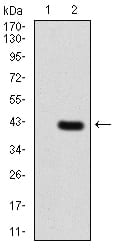
Figure 3:Western blot analysis using PDPK1 mAb against HEK293 (1) and PDPK1 (AA: 457-556)-hIgGFc transfected HEK293 (2) cell lysate.
Western Blot

Figure 4:Western blot analysis using PDPK1 mouse mAb against MCF-7 (1), Hela (2), and U937 (3) cell lysate.
Flow cytometric

Figure 5:Flow cytometric analysis of Hela cells using PDPK1 mouse mAb (green) and negative control (red).
Immunofluorescence analysis

Figure 6:Immunofluorescence analysis of Hela cells using PDPK1 mouse mAb (green). Blue: DRAQ5 fluorescent DNA dye. Red: Actin filaments have been labeled with Alexa Fluor- 555 phalloidin. Secondary antibody from Fisher (Cat#: 35503)
For Research Use Only. Not for use in diagnostic procedures.