
PDK1 Primary Antibody
Item Information
Catalog #
Size
Price
Description
Pyruvate dehydrogenase (PDH) is a mitochondrial multienzyme complex that catalyzes the oxidative decarboxylation of pyruvate and is one of the major enzymes responsible for the regulation of homeostasis of carbohydrate fuels in mammals. The enzymatic activity is regulated by a phosphorylation/dephosphorylation cycle. Phosphorylation of PDH by a specific pyruvate dehydrogenase kinase (PDK) results in inactivation. (provided by RefSeq). Tissue specificity: Expressed predominantly in the heart.
Product Overview
Entrez GenelD
5163
Aliases
PDK1
Clone#
4A11
Host / Isotype
Mouse / IgG1
Species Reactivity
Human, Monkey, Rat
Immunogen
Purified recombinant fragment of human PDK1 expressed in E. Coli.
Formulation
Ascitic fluid containing 0.03% sodium azide.
Storage
Store at 4°C short term. Aliquot and store at -20°C long term. Avoid freeze/thaw cycles.
Product Applications
WB (Western Blot)
1/500 - 1/2000
IHC_P(Immunohistochemistry)
1/200 - 1/1000
ICC (Immunocytochemistry)
1/200 - 1/1000
FCM (Flow Cytometry)
1/200 - 1/400
ELISA
1/10000
References
1. Nat Cell Biol. 2008 Feb;10(2):127-37.
2. Blood. 2008 Apr 1;111(7):3723-34.
3. J Biol Chem. 2007 Apr 20;282(16):12272-89.
2. Blood. 2008 Apr 1;111(7):3723-34.
3. J Biol Chem. 2007 Apr 20;282(16):12272-89.
Product Image
Western Blot

Figure 1: Western blot analysis using PDK1 mouse mAb against NIH/3T3 (1), Hela (2), Jurkat (3), HepG2 (4), PC-12 (5), and Cos7 (6) cell lysate.
Immunohistochemical analysis
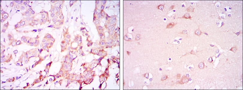
Figure 2: Immunohistochemical analysis of paraffin-embedded breast cancer tissues (left) and brain tissues (right) using PDK1 mouse mAb with DAB staining.
Immunofluorescence analysis

Figure 3: Immunofluorescence analysis of HELA cells using PDK1 mouse mAb (green). Blue: DRAQ5 fluorescent DNA dye. Red: Actin filaments have been labeled with Alexa Fluor-555 phalloidin.
Flow cytometric

Figure 4: Flow cytometric analysis of Lovo cells using PDK1 mouse mAb (green) and negative control (purple).
For Research Use Only. Not for use in diagnostic procedures.