

PDGFRB Primary Antibody
Item Information
Catalog #
Size
Price
Description
This gene encodes a cell surface tyrosine kinase receptor for members of the platelet-derived growth factor family. These growth factors are mitogens for cells of mesenchymal origin. The identity of the growth factor bound to a receptor monomer determines whether the functional receptor is a homodimer or a heterodimer, composed of both platelet-derived growth factor receptor alpha and beta polypeptides. This gene is flanked on chromosome 5 by the genes for granulocyte-macrophage colony-stimulating factor and macrophage-colony stimulating factor receptor; all three genes may be implicated in the 5-q syndrome. A translocation between chromosomes 5 and 12, that fuses this gene to that of the translocation, ETV6, leukemia gene, results in chronic myeloproliferative disorder with eosinophilia.
Product Overview
Entrez GenelD
5159
Aliases
IMF1; IBGC4; JTK12; PDGFR; CD140B; PDGFR1; PDGFR-1
Clone#
2G7B7
Host / Isotype
Mouse / IgG1
Species Reactivity
Human, Mouse
Immunogen
Purified recombinant fragment of human PDGFRB (AA: 33-133) expressed in E. Coli.
Formulation
Purified antibody in PBS with 0.05% sodium azide
Storage
Store at 4°C short term. Aliquot and store at -20°C long term. Avoid freeze/thaw cycles.
Product Applications
WB (Western Blot)
1/500 - 1/2000
FCM (Flow Cytometry)
1/200 - 1/400
ELISA
1/10000
References
1.J Cell Sci. 2012 May 1;125(Pt 9):2276-87.
2.J Cell Physiol. 2012 May;227(5):2089-96.
2.J Cell Physiol. 2012 May;227(5):2089-96.
Product Image
Elisa
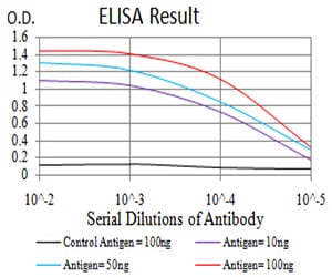
Figure 1: Black line: Control Antigen (100 ng); Purple line: Antigen(10ng); Blue line: Antigen (50 ng); Red line: Antigen (100 ng);
Western Blot

Figure 2:Western blot analysis using PDGFRB mAb against human PDGFRB (AA: 33-133) recombinant protein. (Expected MW is 37 kDa)
Western Blot

Figure 3:Western blot analysis using PDGFRB mAb against HEK293 (1) and PDGFRB (AA: 33-133)-hIgGFc transfected HEK293 (2) cell lysate.
Flow cytometric

Figure 4:Flow cytometric analysis of NIH/3T3 cells using PDGFRB mouse mAb (green) and negative control (red).
For Research Use Only. Not for use in diagnostic procedures.