

PDGFRA Primary Antibody
Item Information
Catalog #
Size
Price
Description
This gene encodes a cell surface tyrosine kinase receptor for members of the platelet-derived growth factor family. These growth factors are mitogens for cells of mesenchymal origin. The identity of the growth factor bound to a receptor monomer determines whether the functional receptor is a homodimer or a heterodimer, composed of both platelet-derived growth factor receptor alpha and beta polypeptides. Studies suggest that this gene plays a role in organ development, wound healing, and tumor progression. Mutations in this gene have been associated with idiopathic hypereosinophilic syndrome, somatic and familial gastrointestinal stromal tumors, and a variety of other cancers.
Product Overview
Entrez GenelD
5156
Aliases
CD140A; PDGFR2; PDGFR-2; RHEPDGFRA
Clone#
8E12F2
Host / Isotype
Mouse / IgG1
Species Reactivity
Human
Immunogen
Purified recombinant fragment of human PDGFRA (AA: 361-528) expressed in E. Coli.
Formulation
Purified antibody from tissue culture in PBS with 0.05% sodium azide
Storage
Store at 4°C short term. Aliquot and store at -20°C long term. Avoid freeze/thaw cycles.
Product Applications
WB (Western Blot)
1/500 - 1/2000
ICC (Immunocytochemistry)
1/200 - 1/1000
FCM (Flow Cytometry)
1/200 - 1/400
ELISA
1/10000
References
1. Stem Cells Dev. 2013 Jul 1;22(13):1932-43.
2. Cancer Biol Ther. 2012 Dec;13(14):1384-9.
2. Cancer Biol Ther. 2012 Dec;13(14):1384-9.
Product Image
Western Blot
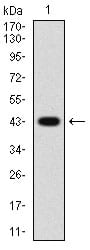
Figure 1: Western blot analysis using PDGFRA mAb against human PDGFRA (AA: 361-528) recombinant protein. (Expected MW is 44.8 kDa)
Western Blot
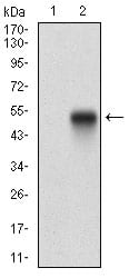
Figure 2: Western blot analysis using PDGFRA mAb against HEK293 (1) and PDGFRA (AA: 361-528)-hIgGFc transfected HEK293 (2) cell lysate.
Immunofluorescence analysis
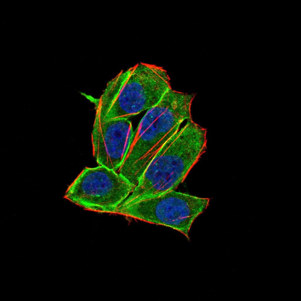
Figure 3:Immunofluorescence analysis of Hela cells using PDGFRA 8E12F2 HELA 100 mouse mAb (green). Blue: DRAQ5 fluorescent DNA dye. Red: Actin filaments have been labeled with Alexa Fluor- 555 phalloidin. Secondary antibody from Fisher (Cat#: 35503)
Flow cytometric
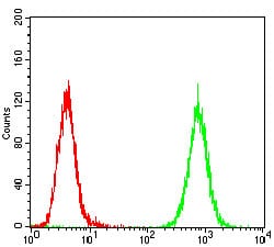
Figure 4: Flow cytometric analysis of Hela cells using PDGFRA mouse mAb (green) and negative control (red).
Elisa
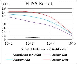
Black line: Control Antigen (100 ng); Purple line: Antigen(10ng); Blue line: Antigen (50 ng); Red line: Antigen (100 ng);
For Research Use Only. Not for use in diagnostic procedures.