
PDE1B Primary Antibody
Item Information
Catalog #
Size
Price
Description
The protein encoded by this gene belongs to the cyclic nucleotide phosphodiesterase (PDE) family, and PDE1 subfamily. Members of the PDE1 family are calmodulin-dependent PDEs that are stimulated by a calcium-calmodulin complex. This PDE has dual-specificity for the second messengers, cAMP and cGMP, with a preference for cGMP as a substrate. cAMP and cGMP function as key regulators of many important physiological processes. Alternatively spliced transcript variants encoding different isoforms have been described for this gene.
Product Overview
Entrez GenelD
5153
Aliases
PDE1B1; PDES1B
Clone#
10B10B12
Host / Isotype
Mouse / IgG1
Species Reactivity
Human, Mouse, Rat
Immunogen
Purified recombinant fragment of human PDE1B (AA: 370-536) expressed in E. Coli.
Formulation
Ascitic fluid containing 0.03% sodium azide.
Storage
Store at 4°C short term. Aliquot and store at -20°C long term. Avoid freeze/thaw cycles.
Product Applications
WB (Western Blot)
1/500 - 1/2000
FCM (Flow Cytometry)
1/200 - 1/400
ELISA
1/10000
References
1.J Biol Chem. 2007 Nov 9;282(45):32749-57.
2.Proc Natl Acad Sci U S A. 2005 Jan 11;102(2):497-502.
2.Proc Natl Acad Sci U S A. 2005 Jan 11;102(2):497-502.
Product Image
Western Blot

Figure 1: Western blot analysis using PDE1B mAb against human PDE1B recombinant protein. (Expected MW is 44.4 kDa)
Western Blot

Figure 2: Western blot analysis using PDE1B mAb against HEK293 (1) and PDE1B (AA: 370-536)-hIgGFc transfected HEK293 (2) cell lysate.
Western Blot
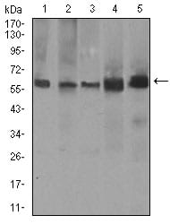
Figure 3: Western blot analysis using PDE1B mouse mAb against A549 (1), SK-MES-1 (2), PC-12 (3), NIH3T3 (4), and 3T3-L1 (5) cell lysate.
Flow cytometric

Figure 4: Flow cytometric analysis of A549 cells using PDE1B mouse mAb (green) and negative control (red).
Elisa

Black line: Control Antigen (100 ng); Purple line: Antigen(10ng); Blue line: Antigen (50 ng); Red line: Antigen (100 ng);
For Research Use Only. Not for use in diagnostic procedures.