

PCNA Primary Antibody
Item Information
Catalog #
Size
Price
Description
The protein encoded by this gene is found in the nucleus and is a cofactor of DNA polymerase delta. The encoded protein acts as a homotrimer and helps increase the processivity of leading strand synthesis during DNA replication. In response to DNA damage, this protein is ubiquitinated and is involved in the RAD6-dependent DNA repair pathway. Two transcript variants encoding the same protein have been found for this gene. Pseudogenes of this gene have been described on chromosome 4 and on the X chromosome.
Product Overview
Entrez GenelD
5111
Clone#
7H4F8
Host / Isotype
Mouse / IgG1
Species Reactivity
Human, Monkey
Immunogen
Purified recombinant fragment of human PCNA (AA: 53-196 ) expressed in E. Coli.
Formulation
Ascitic fluid containing 0.03% sodium azide.
Storage
Store at 4°C short term. Aliquot and store at -20°C long term. Avoid freeze/thaw cycles.
Product Applications
WB (Western Blot)
1/500 - 1/2000
IHC_P(Immunohistochemistry)
1/200 - 1/1000
FCM (Flow Cytometry)
1/200 - 1/400
ELISA
1/10000
References
1.Cancer Res. 2012 Jul 1;72(13):3217-27.
2.PLoS One. 2012;7(1):e29416.
2.PLoS One. 2012;7(1):e29416.
Product Image
Western Blot
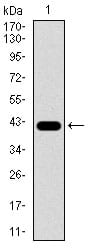
Figure 1: Western blot analysis using PCNA mAb against human PCNA recombinant protein. (Expected MW is 41.2 kDa)
Western Blot

Figure 2: Western blot analysis using PCNA mAb against HEK293 (1) and PCNA (AA: 53-196)-hIgGFc transfected HEK293 (2) cell lysate.
Western Blot

Figure 3: Western blot analysis using PCNA mouse mAb against A431 (1), HEK293 (2), HeLa (3), HepG2 (4), Raji (5), MOLT4 (6), COS7 (7), and MCF-7 (8) cell lysate.
Flow cytometric

Figure 4: Flow cytometric analysis of MOLT4 cells using PCNA mouse mAb (green) and negative control (purple).
Immunohistochemical analysis

Figure 5: Immunohistochemical analysis of paraffin-embedded cervical cancer tissues using PCNA mouse mAb with DAB staining.
Immunohistochemical analysis

Figure 6: Immunohistochemical analysis of paraffin-embedded colon cancer tissues using PCNA mouse mAb with DAB staining.
Elisa

Black line: Control Antigen (100 ng); Purple line: Antigen(10ng); Blue line: Antigen (50 ng); Red line: Antigen (100 ng);
For Research Use Only. Not for use in diagnostic procedures.