

PCK2 Primary Antibody
Item Information
Catalog #
Size
Price
Description
This gene encodes a mitochondrial enzyme that catalyzes the conversion of oxaloacetate to phosphoenolpyruvate in the presence of guanosine triphosphate (GTP). A cytosolic form of this protein is encoded by a different gene and is the key enzyme of gluconeogenesis in the liver. Alternatively spliced transcript variants have been described. [provided by RefSeq, Apr 2014]
Product Overview
Entrez GenelD
5106
Aliases
PEPCK; PEPCK2; PEPCK-M
Clone#
3D3D9
Host / Isotype
Mouse / IgG1
Species Reactivity
Human, Rat, Monkey
Immunogen
Purified recombinant fragment of human PCK2 (AA: 44-175) expressed in E. Coli.
Formulation
Purified antibody in PBS with 0.05% sodium azide
Storage
Store at 4°C short term. Aliquot and store at -20°C long term. Avoid freeze/thaw cycles.
Product Applications
WB (Western Blot)
1/500 - 1/2000
IHC_P(Immunohistochemistry)
1/200 - 1/1000
ICC (Immunocytochemistry)
1/200 - 1/1000
FCM (Flow Cytometry)
1/200 - 1/400
ELISA
1/10000
References
1.Oncogene. 2015 Feb 19;34(8):1044-50.
2.BMC Cancer. 2014 Mar 6;14:160.
2.BMC Cancer. 2014 Mar 6;14:160.
Product Image
Elisa

Figure 1:Black line: Control Antigen (100 ng);Purple line: Antigen (10ng); Blue line: Antigen (50 ng); Red line:Antigen (100 ng)
Western Blot

Figure 2:Western blot analysis using PCK2 mAb against human PCK2 (AA: 44-175) recombinant protein. (Expected MW is 40.5 kDa)
Western Blot

Figure 3:Western blot analysis using PCK2 mAb against HEK293 (1) and PCK2 (AA: 44-175)-hIgGFc transfected HEK293 (2) cell lysate.
Western Blot

Figure 4:Western blot analysis using PCK2 mouse mAb against Jurkat (1), C2C12 (2), Hela (3), HepG2 (4), COS7 (5), and HL-60 (6) cell lysate.
Immunofluorescence analysis

Figure 5:Immunofluorescence analysis of Hela cells using PCK2 mouse mAb (green). Blue: DRAQ5 fluorescent DNA dye. Red: Actin filaments have been labeled with Alexa Fluor- 555 phalloidin. Secondary antibody from Fisher (Cat#: 35503)
Flow cytometric
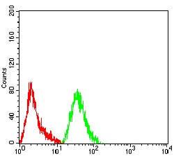
Figure 6:Flow cytometric analysis of Hela cells using PCK2 mouse mAb (green) and negative control (red).
Immunohistochemical analysis

Figure 7:Immunohistochemical analysis of paraffin-embedded stomach cancer tissues using PCK2 mouse mAb with DAB staining.
For Research Use Only. Not for use in diagnostic procedures.