

PAX8 Primary Antibody
Item Information
Catalog #
Size
Price
Description
This gene encodes a member of the paired box (PAX) family of transcription factors. Members of this gene family typically encode proteins that contain a paired box domain, an octapeptide, and a paired-type homeodomain. This nuclear protein is involved in thyroid follicular cell development and expression of thyroid-specific genes. Mutations in this gene have been associated with thyroid dysgenesis, thyroid follicular carcinomas and atypical follicular thyroid adenomas. Alternatively spliced transcript variants encoding different isoforms have been described.
Product Overview
Entrez GenelD
7849
Clone#
1A1D3
Host / Isotype
Mouse / Mouse IgG1
Immunogen
Purified recombinant fragment of human PAX8 (AA: 60-261) expressed in E. Coli.
Formulation
Purified antibody in PBS with 0.05% sodium azide
Storage
Store at 4°C short term. Aliquot and store at -20°C long term. Avoid freeze/thaw cycles.
Product Applications
WB (Western Blot)
1/500 - 1/2000
IHC_P(Immunohistochemistry)
1/200-1/1000
FCM (Flow Cytometry)
1/200-1/400
ELISA
1/10000
References
1.BMC Mol Cell Biol. 2019 Dec 27;20(1):61. 2.PLoS One. 2017 Mar 24;12(3):e0173700.
Product Image
ELISA
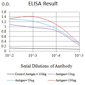
Figure 1: Black line: Control Antigen (100 ng);Purple line: Antigen (10ng); Blue line: Antigen (50 ng); Red line: Antigen (100 ng)
WESTERN BLOT
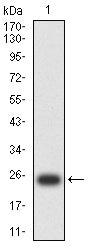
Figure 2: Western blot analysis using PAX8 mAb against human PAX8 (AA: 60-261) recombinant protein. (Expected MW is 25 kDa)
WESTERN BLOT
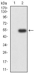
Figure 3: Western blot analysis using PAX8 mAb against HEK293-6e (1) and PAX8 (AA: 60-261)-hIgGFc transfected HEK293 (2) cell lysate.
WESTERN BLOT
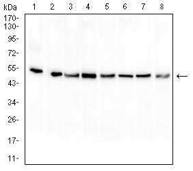
Figure 4: Western blot analysis using PAX8 mouse mAb against HL-60 (1), HEK293 (2), Raji (3), Hela (4), Jurkat (5), A431 (6), A549 (7), and K562 (8) cell lysate.
FLOW CYTOMETRY
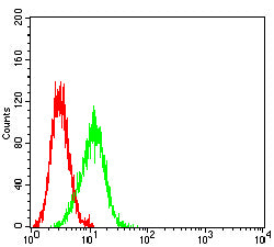
Figure 5: Flow cytometric analysis of SK-OV-3 cells using PAX8 mouse mAb (green) and negative control (red).
IMMUNOHISTOCHEMISTRY

Figure 6: Immunohistochemical analysis of paraffin-embedded kidney tissues using PAX8 mouse mAb with DAB staining.
IMMUNOHISTOCHEMISTRY
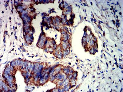
Figure 7: Immunohistochemical analysis of paraffin-embedded rectum cancer tissues using PAX8 mouse mAb with DAB staining.
For Research Use Only. Not for use in diagnostic procedures.