

PAX3 Primary Antibody
Item Information
Catalog #
Size
Price
Description
This gene is a member of the paired box (PAX) family of transcription factors. Members of the PAX family typically contain a paired box domain and a paired-type homeodomain. These genes play critical roles during fetal development. Mutations in paired box gene 3 are associated with Waardenburg syndrome, craniofacial-deafness-hand syndrome, and alveolar rhabdomyosarcoma. The translocation t(2;13)(q35;q14), which represents a fusion between PAX3 and the forkhead gene, is a frequent finding in alveolar rhabdomyosarcoma. Alternative splicing results in transcripts encoding isoforms with different C-termini.
Product Overview
Entrez GenelD
5077
Aliases
WS1; WS3; CDHS; HUP2
Clone#
7D8G7
Host / Isotype
Mouse / IgG1
Species Reactivity
Human, Mouse
Immunogen
Purified recombinant fragment of human PAX3 (AA: 142-203) expressed in E. Coli.
Formulation
Purified antibody in PBS with 0.05% sodium azide
Storage
Store at 4°C short term. Aliquot and store at -20°C long term. Avoid freeze/thaw cycles.
Product Applications
WB (Western Blot)
1/500 - 1/2000
ELISA
1/10000
References
1. Cell Death Differ. 2012 Apr;19(4):616-22.
2. Biochem Biophys Res Commun. 2011 Aug 12;411(4):832-7.
2. Biochem Biophys Res Commun. 2011 Aug 12;411(4):832-7.
Product Image
Western Blot
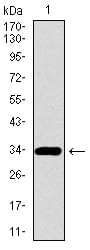
Figure 1: Western blot analysis using PAX3 mAb against human PAX3 recombinant protein. (Expected MW is 32.6 kDa)
Western Blot
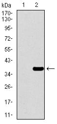
Figure 2: Western blot analysis using PAX3 mAb against HEK293 (1) and PAX3 (AA: 142-203)-hIgGFc transfected HEK293 (2) cell lysate.
Western Blot
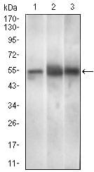
Figure 3: Western blot analysis using PAX3 mouse mAb against Mouse brain (1), Rat spleen (2), Mouse liver (3) cell lysate.
Elisa
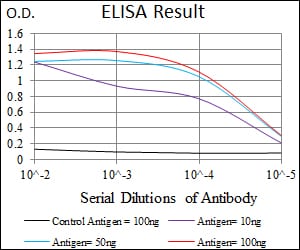
Black line: Control Antigen (100 ng); Purple line: Antigen(10ng); Blue line: Antigen (50 ng); Red line: Antigen (100 ng);
For Research Use Only. Not for use in diagnostic procedures.