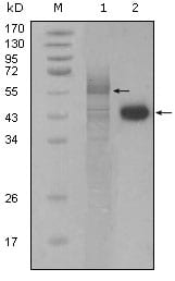
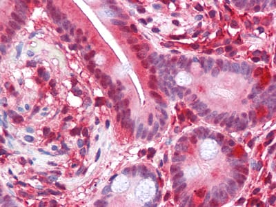
PAR4 Primary Antibody

Item Information
Catalog #
Size
Price
Description

Prostate apoptosis response 4 (Par4) is a 38kD protein originally identified as the product of a gene that is upregulated in prostate tumor cells undergoing apoptosis. It is a leucine zipper and death domain containing protein whose levels increase in neurons undergoing apoptosis as a result of trophic factor withdrawal or exposure to oxidative and metabolic insults. Par4 levels are reported to be increased in their lumbar spinal cord specimens further suggesting a role in neuronal degeneration. The tumor suppressor WT1 represses and activates transcription. The loss and/or imbalance of the dual transcriptional activity of WT1 may contribute to Wilms tumor. Par4 is a WT1 interacting protein that also functions as a transcriptional repressor.

Product Overview
Entrez GenelD
5074
Aliases
PAWR
Clone#
3G9H7
Host / Isotype
Mouse / IgG1
Species Reactivity
Human
Immunogen
Purified recombinant fragment of PAR4(aa1-330) expressed in E. Coli.
Formulation
Ascitic fluid containing 0.03% sodium azide.
Storage
4°C; -20°C for long term storage
Product Applications
WB (Western Blot)
1/500 - 1/2000
IHC_P(Immunohistochemistry)
1/200 - 1/1000
ELISA
1/10000
References
1. J Biol Chem. 2004 Jul 2;279(27):28266-75.
2. Exp Hematol. 2004 Jul;32(7):649-56.
3. Mol Cell Biol. 2005 Feb;25(3):1146-61.
4. Psychiatr Genet. 2006 Oct;16(5):193-6.
Product Image
Western Blot
Figure 1: Western blot analysis using PAR4 mouse mAb against full-length Trx-Par4 recombinant protein (1) and Hela cell lysate (2).
Immunohistochemical analysis
Figure 2: Immunohistochemical analysis of paraffin-embedded human brain (left) and breast carcinoma (right), showing cytoplasmic and membrane localization using PAR4 mouse mAb with DAB staining.
Immunohistochemical analysis
Figure 3: Immunohistochemical analysis of paraffin-embedded human Small Intestine tissues using PAR4 mouse mAb
For Research Use Only. Not for use in diagnostic procedures.