
PAPLN Primary Antibody
Item Information
Catalog #
Size
Price
Description
Papilin is an extracellular matrix glycoprotein involved in, thin matrix layers during gastrulation, matrix associated with wandering, phagocytic hemocytes, basement membranes and space-filling matrix during Drosophila development.Determination of its cDNA sequence led to the identification of Caenorhabditis and mammalian papilins. A distinctly conserved 'papilin cassette' of domains at the amino-end of papilins is also the carboxyl-end of the ADAMTS subgroup of secreted, matrix-associated metalloproteinases; this cassette contains one thrombospondin type 1 (TSR) domain, a specific cysteine-rich domain and several partial TSR domains. In vitro, papilin non-competitively inhibits procollagen N-proteinase, an ADAMTS metalloproteinase.
Product Overview
Entrez GenelD
89932
Aliases
Papilin
Clone#
5B2E5
Host / Isotype
Mouse / IgG1
Species Reactivity
Human, Mouse
Immunogen
Purified recombinant fragment of human PAPLN (AA: 766-870) expressed in E. Coli.
Formulation
Purified antibody in PBS with 0.05% sodium azide
Storage
Store at 4°C short term. Aliquot and store at -20°C long term. Avoid freeze/thaw cycles.
Product Applications
WB (Western Blot)
1/500 - 1/2000
ICC (Immunocytochemistry)
1/200 - 1/1000
ELISA
1/10000
References
1. Int J Biochem Cell Biol. 2004 Jun; 36(6):1079-84.
2. Development. 2000 Dec;127(24):5475-85.
2. Development. 2000 Dec;127(24):5475-85.
Product Image
Western Blot

Figure 1: Western blot analysis using PAPLN mAb against human PAPLN recombinant protein. (Expected MW is 36.4 kDa)
Western Blot

Figure 2: Western blot analysis using PAPLN mAb against HEK293 (1) and PAPLN (AA: 766-870)-hIgGFc transfected HEK293 (2) cell lysate.
Western Blot

Figure 3: Western blot analysis using PAPLN mouse mAb against Hela (1), HepG2 (2), OCM-1 (3), Raji (4), Jurkat (5), NIH/3T3 (6) cell lysate.
Immunofluorescence analysis
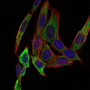
Figure 4: Immunofluorescence analysis of NIH/3T3 cells using PAPLN mouse mAb (green). Blue: DRAQ5 fluorescent DNA dye. Red: Actin filaments have been labeled with Alexa Fluor-555 phalloidin.
Elisa

Black line: Control Antigen (100 ng); Purple line: Antigen(10ng); Blue line: Antigen (50 ng); Red line: Antigen (100 ng);
For Research Use Only. Not for use in diagnostic procedures.