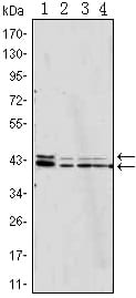
p44/42 MAPK (Erk1/2) Primary Antibody

Item Information
Catalog #
Size
Price
Description

Mitogen-activated protein kinases (MAPKs) are a widely conserved family of serine/threonine protein kinases involved in many cellular programs such as cell proliferation, differentiation, motility, and death. The p44/42 MAPK (Erk1/2) signaling pathway can be activated in response to a diverse range of extracellular stimuli including mitogens, growth factors, and cytokines and is an important target in the diagnosis and treatment of cancer. Upon stimulation, a sequential three-part protein kinase cascade is initiated, consisting of a MAP kinase kinase kinase (MAPKKK or MAP3K), a MAP kinase kinase (MAPKK or MAP2K), and a MAP kinase (MAPK). Multiple p44/42 MAP3Ks have been identified, including members of the Raf family as well as Mos and Tpl2/Cot. MEK1 and MEK2 are the primary MAPKKs in this pathway. MEK1 and MEK2 activate p44 and p42 through phosphorylation of activation loop residues Thr202/Tyr204 and Thr185/Tyr187, respectively. Several downstream targets of p44/42 have been identified, including p90RSK and the transcription factor Elk-1. p44/42 are negatively regulated by a family of dual-specificity (Thr/Tyr) MAPK phosphatases, known as DUSPs or MKPs, along with MEK inhibitors such as U0126 and PD98059.

Product Overview
Entrez GenelD
5594
Aliases
ERK; p38; p40; p41; ERK2; ERT1; MAPK2; PRKM1; PRKM2; P42MAPK; p41mapk; MAPK1
Clone#
3F8
Host / Isotype
Mouse / IgG2b
Species Reactivity
Human, Mouse
Immunogen
Purified recombinant fragment of human MAPK expressed in E. Coli.
Formulation
Ascitic fluid containing 0.03% sodium azide.
Storage
4°C; -20°C for long term storage
Product Applications
WB (Western Blot)
1/500 - 1/2000
IHC_P(Immunohistochemistry)
1/200 - 1/1000
FCM (Flow Cytometry)
1/200 - 1/400
ELISA
1/10000
References
1. FEBS Lett. 1992 Jun 15;304(2-3):170-8.
2. Proc Natl Acad Sci U S A. 1995 Jun 6;92(12):5361-5.
3. J Biol Chem. 2009 Nov 27;284(48):33456-65.
4. BMC Biol. 2009 Oct 27;7:70.
Product Image
Western Blot
Figure 1: Western blot analysis using p44/42 MAPK mouse mAb against Jurkat (1), Hela (2), A431 (3) and NIH/3T3 (4) cell lysate.
Immunohistochemical analysis
Figure 2: Immunohistochemical analysis of paraffin-embedded human Liver tissues using anti-BHMT mouse mAb
Flow cytometric
Figure 3: Flow cytometric analysis of Jurkat cells using p44/42 MAPK mAb (green) and negative control (purple).
For Research Use Only. Not for use in diagnostic procedures.