

P2RY14 Primary Antibody
Item Information
Catalog #
Size
Price
Description
The product of this gene belongs to the family of G-protein coupled receptors, which contains several receptor subtypes with different pharmacological selectivity for various adenosine and uridine nucleotides. This receptor is a P2Y purinergic receptor for UDP-glucose and other UDP-sugars coupled to G-proteins. It has been implicated in extending the known immune system functions of P2Y receptors by participating in the regulation of the stem cell compartment, and it may also play a role in neuroimmune function. Two transcript variants encoding the same protein have been identified for this gene.
Product Overview
Entrez GenelD
9934
Aliases
P2Y14; BPR105; GPR105
Clone#
8A11B11
Host / Isotype
Mouse / IgG2b
Species Reactivity
Human
Immunogen
Purified recombinant fragment of human P2RY14 (AA: extra mix) expressed in E. Coli.
Formulation
Purified antibody in PBS with 0.05% sodium azide
Storage
Store at 4°C short term. Aliquot and store at -20°C long term. Avoid freeze/thaw cycles.
Product Applications
WB (Western Blot)
1/500 - 1/2000
IHC_P(Immunohistochemistry)
1/200 - 1/1000
FCM (Flow Cytometry)
1/200 - 1/400
ELISA
1/10000
References
1.Mol Pharmacol. 2013 Jul;84(1):41-9.
2.Bioorg Med Chem Lett. 2007 Feb 1;17(3):761-6.
2.Bioorg Med Chem Lett. 2007 Feb 1;17(3):761-6.
Product Image
Elisa

Figure 1: Black line: Control Antigen (100 ng);Purple line: Antigen (10ng); Blue line: Antigen (50 ng); Red line:Antigen (100 ng)
Western Blot

Figure 2:Western blot analysis using P2RY14 mAb against human P2RY14 (AA: extra mix) recombinant protein. (Expected MW is 11.5 kDa)
Western Blot
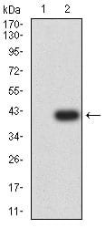
Figure 3:Western blot analysis using P2RY14 mAb against HEK293 (1) and P2RY14 (AA: extra mix)-hIgGFc transfected HEK293 (2) cell lysate.
Flow cytometric

Figure 4:Flow cytometric analysis of Hela cells using P2RY14 mouse mAb (green) and negative control (red).
Immunohistochemical analysis
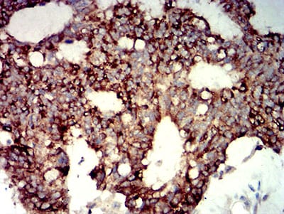
Figure 5:Immunohistochemical analysis of paraffin-embedded colon cancer tissues using P2RY14 mouse mAb with DAB staining.
Immunohistochemical analysis
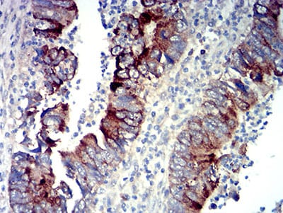
Figure 6:Immunohistochemical analysis of paraffin-embedded rectum cancer tissues using P2RY14 mouse mAb with DAB staining.
For Research Use Only. Not for use in diagnostic procedures.