

P2RY1 Primary Antibody
Item Information
Catalog #
Size
Price
Description
The product of this gene belongs to the family of G-protein coupled receptors. This family has several receptor subtypes with different pharmacological selectivity, which overlaps in some cases, for various adenosine and uridine nucleotides. This receptor functions as a receptor for extracellular ATP and ADP. In platelets binding to ADP leads to mobilization of intracellular calcium ions via activation of phospholipase C, a change in platelet shape, and probably to platelet aggregation.
Product Overview
Entrez GenelD
5028
Aliases
P2Y1
Clone#
4G5D6
Host / Isotype
Mouse / IgG2b
Species Reactivity
Human, Rat
Immunogen
Purified recombinant fragment of human P2RY1 (AA: extra mix) expressed in E. Coli.
Formulation
Purified antibody in PBS with 0.05% sodium azide
Storage
Store at 4°C short term. Aliquot and store at -20°C long term. Avoid freeze/thaw cycles.
Product Applications
WB (Western Blot)
1/500 - 1/2000
IHC_P(Immunohistochemistry)
1/200 - 1/1000
FCM (Flow Cytometry)
1/200 - 1/400
ELISA
1/10000
References
1.Nature. 2015 Apr 16;520(7547):317-21.
2.Biochem Pharmacol. 2011 Aug 15;82(4):418-25.
2.Biochem Pharmacol. 2011 Aug 15;82(4):418-25.
Product Image
Elisa
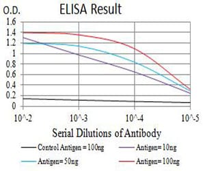
Figure 1: Black line: Control Antigen (100 ng);Purple line: Antigen (10ng); Blue line: Antigen (50 ng); Red line:Antigen (100 ng)
Western Blot
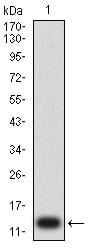
Figure 2:Western blot analysis using P2RY1 mAb against human P2RY1 (AA: extra mix) recombinant protein. (Expected MW is 13 kDa)
Western Blot
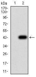
Figure 3:Western blot analysis using P2RY1 mAb against HEK293 (1) and P2RY1 (AA: extra mix)-hIgGFc transfected HEK293 (2) cell lysate.
Western Blot
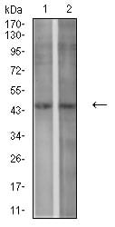
Figure 4:Western blot analysis using P2RY1 mouse mAb against SPC-A-1 (1) and C6 (2) cell lysate.
Flow cytometric
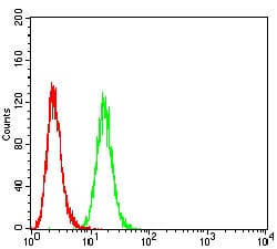
Figure 5:Flow cytometric analysis of Hela cells using P2RY1 mouse mAb (green) and negative control (red).
Flow cytometric
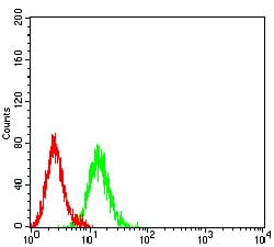
Figure 6:Flow cytometric analysis of K562 cells using P2RY1 mouse mAb (green) and negative control (red).
Immunohistochemical analysis
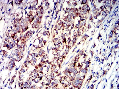
Figure 7:Immunohistochemical analysis of paraffin-embedded bladder cancer tissues using P2RY1 mouse mAb with DAB staining.
Immunohistochemical analysis

Figure 8:Immunohistochemical analysis of paraffin-embedded rectum cancer tissues using P2RY1 mouse mAb with DAB staining.
For Research Use Only. Not for use in diagnostic procedures.