
OTX2 Primary Antibody
Item Information
Catalog #
Size
Price
Description
This gene encodes a member of the bicoid sub-family of homeodomain-containing transcription factors. The encoded protein acts as a transcription factor and may play a role in brain and sensory organ development. A similar protein in mice is required for proper forebrain development.Tissue specificity: Expressed in brain.
Product Overview
Entrez GenelD
5015
Aliases
MCOPS5; MGC45000
Clone#
1H12C4B5
Host / Isotype
Mouse / IgG1
Species Reactivity
Human
Immunogen
Purified recombinant fragment of human OTX2 expressed in E. Coli.
Formulation
Ascitic fluid containing 0.03% sodium azide.
Storage
Store at 4°C short term. Aliquot and store at -20°C long term. Avoid freeze/thaw cycles.
Product Applications
WB (Western Blot)
1/500 - 1/2000
IHC_P(Immunohistochemistry)
1/200 - 1/1000
ICC (Immunocytochemistry)
1/200 - 1/1000
FCM (Flow Cytometry)
1/200 - 1/400
ELISA
1/10000
References
1. Hum Mutat. 2008 Nov;29(11):E278-83.
2. Cancer Res. 2010 Jan 1;70(1):181-91.
2. Cancer Res. 2010 Jan 1;70(1):181-91.
Product Image
Western Blot

Figure 1: Western blot analysis using OTX2 mouse mAb against HepG2 (1), Jurkat (2), and NTERA-2 (3) cell lysate.
Western Blot

Figure 2: Western blot analysis using OTX2 mAb against human OTX2 (AA: 40-297) recombinant protein. (Expected MW is 65 kDa)
Immunohistochemical analysis
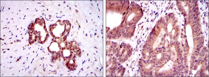
Figure 3: Immunohistochemical analysis of paraffin-embedded prostate tissues (left) and colon cancer tissues (right) using OTX2 mouse mAb with DAB staining.
Immunofluorescence analysis

Figure 4: Immunofluorescence analysis of HepG2 cells using OTX2 mouse mAb (green). Red: Actin filaments have been labeled with Alexa Fluor-555 phalloidin.
Flow cytometric

Figure 5: Flow cytometric analysis of HepG2 cells using OTX2 mouse mAb (green) and negative control (purple).
Elisa

Red: Control Antigen (100ng); Purple: Antigen (10ng); Green: Antigen (50ng); Blue: Antigen (100ng);
For Research Use Only. Not for use in diagnostic procedures.