

NT5E Primary Antibody
Item Information
Catalog #
Size
Price
Description
The protein encoded by this gene is a plasma membrane protein that catalyzes the conversion of extracellular nucleotides to membrane-permeable nucleosides. The encoded protein is used as a determinant of lymphocyte differentiation. Defects in this gene can lead to the calcification of joints and arteries. Two transcript variants encoding different isoforms have been found for this gene.
Product Overview
Entrez GenelD
4907
Aliases
NT; eN; NT5; NTE; eNT; CD73; E5NT; CALJA
Clone#
4G6B10
Host / Isotype
Mouse / IgG1
Species Reactivity
Human
Immunogen
Purified recombinant fragment of human NT5E (AA: 30-250) expressed in E. Coli.
Formulation
Purified antibody in PBS with 0.05% sodium azide.
Storage
Store at 4°C short term. Aliquot and store at -20°C long term. Avoid freeze/thaw cycles.
Product Applications
WB (Western Blot)
1/500 - 1/2000
IHC_P(Immunohistochemistry)
1/200 - 1/1000
ELISA
1/10000
References
1. Appl Immunohistochem Mol Morphol. 2012 Mar;20(2):103-7.
2. J Surg Oncol. 2012 Aug 1;106(2):130-7.
2. J Surg Oncol. 2012 Aug 1;106(2):130-7.
Product Image
Western Blot

Figure 1: Western blot analysis using NT5E mAb against human NT5E (AA: ) recombinant protein. (Expected MW is 26.6 kDa)
Western Blot

Figure 2: Western blot analysis using NT5E mAb against HEK293 (1) and NT5E (AA: 30-250)-hIgGFc transfected HEK293 (2) cell lysate.
Western Blot

Figure 3: Western blot analysis using NT5E mouse mAb against A431 (1) cell lysate.
Immunohistochemical analysis
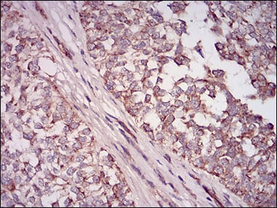
Figure 4: Immunohistochemical analysis of paraffin-embedded bladder cancer tissues using NT5E mouse mAb with DAB staining.
Immunohistochemical analysis

Figure 5: Immunohistochemical analysis of paraffin-embedded esophagus cancer tissues using NT5E mouse mAb with DAB staining.
Elisa

Black line: Control Antigen (100 ng); Purple line: Antigen(10ng); Blue line: Antigen (50 ng); Red line: Antigen (100 ng);
For Research Use Only. Not for use in diagnostic procedures.