
NR3C1 Primary Antibody
Item Information
Catalog #
Size
Price
Description
The protein encoded by this gene is a receptor for glucocorticoids that can act as both a transcription factor and as a regulator of other transcription factors. This protein can also be found in heteromeric cytoplasmic complexes along with heat shock factors and immunophilins. The protein is typically found in the cytoplasm until it binds a ligand, which induces transport into the nucleus. Mutations in this gene are a cause of glucocorticoid resistance, or cortisol, resistance. Tissue specificity: Widely expressed. In the heart, detected in left and right atria, left and right ventricles, aorta, apex, intraventricular septum, and atrioventricular node as well as whole adult and fetal heart.
Product Overview
Entrez GenelD
2908
Aliases
GR; GCR; GRL; GCCR; NR3C1
Clone#
6E6
Host / Isotype
Mouse / IgG1
Species Reactivity
Human
Immunogen
Purified recombinant fragment of human NR3C1 expressed in E. Coli.
Formulation
Ascitic fluid containing 0.03% sodium azide.
Storage
Store at 4°C short term. Aliquot and store at -20°C long term. Avoid freeze/thaw cycles.
Product Applications
WB (Western Blot)
1/500 - 1/2000
IHC_P(Immunohistochemistry)
1/200 - 1/1000
ICC (Immunocytochemistry)
1/200 - 1/1000
FCM (Flow Cytometry)
1/200 - 1/400
ELISA
1/10000
References
1. J Clin Endocrinol Metab. 2008 Dec;93(12):4963-8.
2. Epigenetics. 2008 Mar-Apr;3(2):97-106.
2. Epigenetics. 2008 Mar-Apr;3(2):97-106.
Product Image
Western Blot

Figure 1: Western blot analysis using NR3C1 mouse mAb against Hela cell lysate.
Immunohistochemical analysis

Figure 2: Immunohistochemical analysis of paraffin-embedded lung cancer tissues using NR3C1 mouse mAb with DAB staining.
Immunofluorescence analysis

Figure 3: Immunofluorescence analysis of PC-2 cells using NR3C1 mouse mAb (green). Blue: DRAQ5 fluorescent DNA dye. Red: Actin filaments have been labeled with Alexa Fluor-555 phalloidin.
Flow cytometric
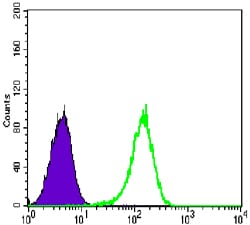
Figure 4: Flow cytometric analysis of K562 cells using NR3C1 mouse mAb (green) and negative control (purple).
Elisa
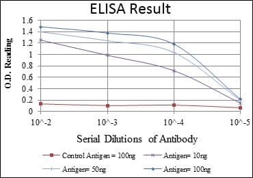
Red: Control Antigen (100ng); Purple: Antigen (10ng); Green: Antigen (50ng); Blue: Antigen (100ng);
For Research Use Only. Not for use in diagnostic procedures.