
Mouse Monoclonal Antibody to NR1H2
Item Information
Catalog #
Size
Price
Description
The liver X receptors, LXRA (NR1H3; MIM 602423) and LXRB, form a subfamily of the nuclear receptor superfamily and are key regulators of macrophage function, controlling transcriptional programs involved in lipid homeostasis and inflammation. The inducible LXRA is highly expressed in liver, adrenal gland, intestine, adipose tissue, macrophages, lung, and kidney, whereas LXRB is ubiquitously expressed. Ligand-activated LXRs form obligate heterodimers with retinoid X receptors (RXRs; see MIM 180245) and regulate expression of target genes containing LXR response elements (summary by Korf et al., 2009 [PubMed 19436111]).
Product Overview
Entrez GenelD
7376
Aliases
NER; UNR; LXRB; LXR-b; NER-I; RIP15
Clone#
5C11D3
Host / Isotype
Mouse / IgG2b
Immunogen
Purified recombinant fragment of human NR1H2 (AA:1-200) expressed in E. Coli.
Formulation
Purified antibody in PBS with 0.05% sodium azide
Storage
Store at 4°C short term. Aliquot and store at -20°C long term. Avoid freeze/thaw cycles.
Product Applications
WB (Western Blot)
1/500 - 1/2000
ICC (Immunocytochemistry)
1/200 - 1/1000
FCM (Flow Cytometry)
1/200 - 1/400
ELISA
1/10000
References
1,J Cell Mol Med. 2019 Feb;23(2):789-797. 2,Biochim Biophys Acta Mol Cell Biol Lipids. 2018 Sep;1863(9):968-979.
Product Image
Elisa
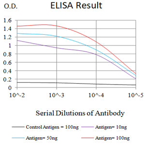
Figure 1:Black line: Control Antigen (100 ng);Purple line: Antigen (10ng); Blue line: Antigen (50 ng); Red line:Antigen (100 ng)
Western Blot

Figure 2:Western blot analysis using NR1H2 mAb against human NR1H2 (AA:1-200) recombinant protein. (Expected MW is 24.4 kDa)
Western Blot

Figure 3:Western blot analysis using NR1H2 mAb against HEK293-6e (1) and human NR1H2 (AA:1-200)-hIgGFc transfected HEK293-6e (2) cell lysate.
Flow cytometric analysis

Figure 4:Flow cytometric analysis of A375 cells using NR1H2 mouse mAb (green) and negative control (red).
Flow cytometric analysis

Figure 5:Flow cytometric analysis of Hela cells using NR1H2 mouse mAb (green) and negative control (red).
Flow cytometric analysis

Figure 6:Flow cytometric analysis of Raji cells using NR1H2 mouse mAb (green) and negative control (red).
Immunofluorescence analysis

Figure 7:Immunofluorescence analysis of Hela cells using NR1H2 mouse mAb (green). Blue: DRAQ5 fluorescent DNA dye. Red: Actin filaments have been labeled with Alexa Fluor- 555 phalloidin. Secondary antibody from Fisher (Cat#: 35503)
For Research Use Only. Not for use in diagnostic procedures.