

NOS2 Primary Antibody
Item Information
Catalog #
Size
Price
Description
Nitric oxide is a reactive free radical which acts as a biologic mediator in several processes, including neurotransmission and antimicrobial and antitumoral activities. This gene encodes a nitric oxide synthase which is expressed in liver and is inducible by a combination of lipopolysaccharide and certain cytokines. Three related pseudogenes are located within the Smith-Magenis syndrome region on chromosome 17.
Product Overview
Entrez GenelD
4843
Aliases
NOS; INOS; NOS2A; HEP-NOS
Clone#
4E5
Host / Isotype
Mouse / IgG1
Species Reactivity
Human, Mouse
Immunogen
Purified recombinant fragment of human NOS2 expressed in E. Coli.
Formulation
Purified antibody in PBS with 0.05% sodium azide
Storage
Store at 4°C short term. Aliquot and store at -20°C long term. Avoid freeze/thaw cycles.
Product Applications
WB (Western Blot)
1/500 - 1/2000
IHC_P(Immunohistochemistry)
1/200 - 1/1000
FCM (Flow Cytometry)
1/200 - 1/400
ELISA
1/10000
References
1. Pediatr Allergy Immunol. 2010 Dec;21(8):1151-6.
2. J Biol Chem. 2010 Dec 31;285(53):41422-31.
2. J Biol Chem. 2010 Dec 31;285(53):41422-31.
Product Image
Western Blot

Figure 1: Western blot analysis using NOS2 mAb against human NOS2 (AA: 997-1058) recombinant protein. (Expected MW is 32.6 kDa)
Western Blot

Figure 2: Western blot analysis using NOS2 mouse mAb against Jurkat (1), Jurkat (2), A549 (3), HeLa (4), NIH3T3 (5)and MCF-7 (6) cell lysate.
Immunohistochemical analysis

Figure 3: Immunohistochemical analysis of paraffin-embedded liver cancer tissues using NOS2 mouse mAb with DAB staining.
Immunohistochemical analysis
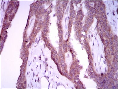
Figure 4: Immunohistochemical analysis of paraffin-embedded breast cancer tissues using NOS2 mouse mAb with DAB staining.
Flow cytometric

Figure 5: Flow cytometric analysis of MCF-7 cells using NOS2 mouse mAb (green) and negative control (red).
Elisa

Black line: Control Antigen (100 ng); Purple line: Antigen(10ng); Blue line: Antigen (50 ng); Red line: Antigen (100 ng);
For Research Use Only. Not for use in diagnostic procedures.