
NME2 Primary Antibody
Item Information
Catalog #
Size
Price
Description
Nucleoside diphosphate kinase (NDK) exists as a hexamer composed of 'A' (encoded by NME1) and 'B' (encoded by this gene) isoforms. Multiple alternatively spliced transcript variants have been found for this gene. Read-through transcription from the neighboring upstream gene (NME1) generates naturally-occurring transcripts (NME1-NME2) that encode a fusion protein comprised of sequence sharing identity with each individual gene product.
Product Overview
Entrez GenelD
4831
Aliases
PUF; NDKB; NDPKB; NM23B; NDPK-B; NM23-H2
Clone#
4G7A8
Host / Isotype
Mouse / IgG1
Species Reactivity
Human
Immunogen
Purified recombinant fragment of human NME2 (AA: FULL(1-152)) expressed in E. Coli.
Formulation
Purified antibody in PBS with 0.05% sodium azide.
Storage
Store at 4°C short term. Aliquot and store at -20°C long term. Avoid freeze/thaw cycles.
Product Applications
WB (Western Blot)
1/500 - 1/2000
IHC_P(Immunohistochemistry)
1/200 - 1/1000
ELISA
1/10000
References
1. Carcinogenesis. 2011 Aug;32(8):1133-42.
2. Cancer Lett. 2009 Mar 18;275(2):221-6.
2. Cancer Lett. 2009 Mar 18;275(2):221-6.
Product Image
Western Blot

Figure 1: Western blot analysis using NME2 mAb against human NME2 (AA: FULL(1-152)) recombinant protein. (Expected MW is 43.2 kDa)
Western Blot

Figure 2: Western blot analysis using NME2 mAb against HEK293 (1) and NME2 (AA: FULL(1-152))-hIgGFc transfected HEK293 (2) cell lysate.
Immunohistochemical analysis

Figure 3: Immunohistochemical analysis of paraffin-embedded ovarian cancer tissues using NME2 mouse mAb with DAB staining.
Western Blot

Figure 4:Western blot analysis using NME2 mouse mAb against Hela (1), and Raji (2) cell lysate.
Elisa
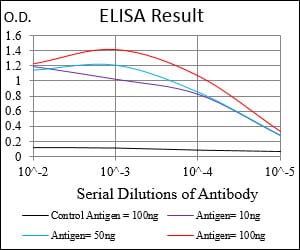
Black line: Control Antigen (100 ng); Purple line: Antigen(10ng); Blue line: Antigen (50 ng); Red line: Antigen (100 ng);
For Research Use Only. Not for use in diagnostic procedures.