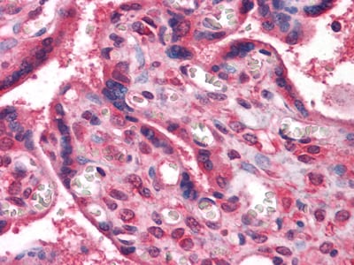
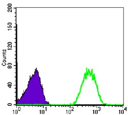
NME1 Primary Antibody
non-metastatic cells 1,protein,with a nm23 nucleoside diphosphate kinase gene family,involved in the phosphorylation of nucleoside diphosphates,with a reduced expression in tumor progression to the metastatic phenotype,mutated in agressive neuroblastoma,expressed in lung carcinoma cell lines not in normal lung,pyrimidine biosynthetic pathway. Involved in cell proliferation, differentiation and development, signal transduction, G protein-coupled receptor endocytosis, and gene expression. Required for neural development including neural patterning and cell fate determination. Has tumor metastasis-suppressive capacity.Tissue specificity: Isoform 1 is expressed in heart, brain, placenta, lung, liver, skeletal muscle, pancreas, spleen and thymus. Expressed in lung carcinoma cell lines but not in normal lung tissues. Isoform 2 is ubiquitously expressed and its expression is also related to tumor differentiation. Isoform 3 is ubiquitously expressed.
2. FEBS Lett. 2009 Sep 3;583(17):2789-92.
3. PLoS One. 2009 Nov 23;4(11):e7949.