

Mouse Monoclonal Antibody to NGFR
Item Information
Catalog #
Size
Price
Description
Nerve growth factor receptor contains an extracellular domain containing four 40-amino acid repeats with 6 cysteine residues at conserved positions followed by a serine/threonine-rich region, a single transmembrane domain, and a 155-amino acid cytoplasmic domain. The cysteine-rich region contains the nerve growth factor binding domain.
Product Overview
Entrez GenelD
4804
Aliases
CD271; p75NTR; TNFRSF16; p75(NTR); Gp80-LNGFR
Clone#
8G8H10
Host / Isotype
Mouse / IgG1
Immunogen
Purified recombinant fragment of human NGFR (AA: 1-200 ) expressed in E. Coli.
Formulation
Purified antibody in PBS with 0.05% sodium azide
Storage
Store at 4°C short term. Aliquot and store at -20°C long term. Avoid freeze/thaw cycles.
Product Applications
WB (Western Blot)
1/500 - 1/2000
IHC_P(Immunohistochemistry)
1/200 - 1/1000
FCM (Flow Cytometry)
1/200 - 1/400
ELISA
1/10000
References
1.Math Biosci Eng. 2019 Sep 5;16(6):8060-8068. 2.Sci Rep. 2019 May 22;9(1):7696.
Product Image
Elisa
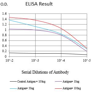
Figure 1:Black line: Control Antigen (100 ng);Purple line: Antigen (10ng); Blue line: Antigen (50 ng); Red line:Antigen (100 ng)
Western Blot

Figure 2:Western blot analysis using NGFR mAb against human NGFR (AA: 1-200) recombinant protein. (Expected MW is 32.7 kDa)
Western Blot

Figure 3:Western blot analysis using NGFR mAb against HEK293-6e (1) and NGFR (AA: 1-200)-hIgGFc transfected HEK293-6e (2) cell lysate.
Flow cytometric analysis

Figure 4:Flow cytometric analysis of Hela cells using NGFR mouse mAb (green) and negative control (red).
Immunohistochemical analysis

Figure 5:Immunohistochemical analysis of paraffin-embedded cerebrum tissues using NGFR mouse mAb with DAB staining.
Immunohistochemical analysis

Figure 6:Immunohistochemical analysis of paraffin-embedded cervical cancer tissues using NGFR mouse mAb with DAB staining.
For Research Use Only. Not for use in diagnostic procedures.