

NCK1 Primary Antibody
Item Information
Catalog #
Size
Price
Description
The protein encoded by this gene is one of the signaling and transforming proteins containing Src homology 2 and 3 (SH2 and SH3) domains. It is located in the cytoplasm and is an adaptor protein involved in transducing signals from receptor tyrosine kinases to downstream signal recipients such as RAS. Alternatively spliced transcript variants encoding different isoforms have been found.
Product Overview
Entrez GenelD
4690
Aliases
NCK; nck-1; NCKalpha
Clone#
5B7
Host / Isotype
Mouse / IgG1
Species Reactivity
Human, Monkey
Immunogen
Purified recombinant fragment of human NCK1 expressed in E. Coli.
Formulation
Purified antibody in PBS with 0.05% sodium azide
Storage
Store at 4°C short term. Aliquot and store at -20°C long term. Avoid freeze/thaw cycles.
Product Applications
WB (Western Blot)
1/500 - 1/2000
IHC_P(Immunohistochemistry)
1/200 - 1/1000
FCM (Flow Cytometry)
1/200 - 1/400
ELISA
1/10000
References
1. Mol Cell Biol. 2008 Mar;28(6):2035-46.
2. Cell Signal. 2010 May;22(5):848-56.
2. Cell Signal. 2010 May;22(5):848-56.
Product Image
Western Blot
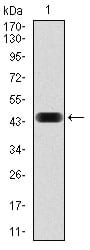
Figure 1: Western blot analysis using NCK1 mAb against human NCK1 (AA: 203-371) recombinant protein. (Expected MW is 44.9 kDa)
Western Blot

Figure 2: Western blot analysis using NCK1 mouse mAb against Jurkat (1), HeLa (2), HEK293 (3), A431 (4), K562 (5), and COS7 (6) cell lysate.
Immunohistochemical analysis
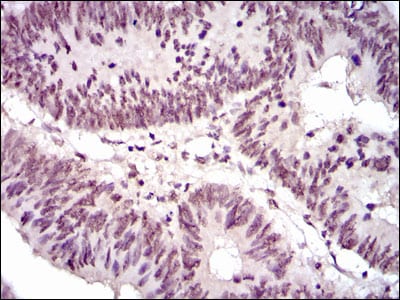
Figure 3: Immunohistochemical analysis of paraffin-embedded rectum cancer tissues using NCK1 mouse mAb with DAB staining.
Flow cytometric

Figure 4: Flow cytometric analysis of Jurkat cells using NCK1 mouse mAb (green) and negative control (red).
Elisa
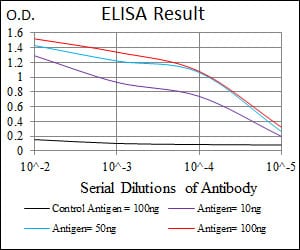
Black line: Control Antigen (100 ng); Purple line: Antigen(10ng); Blue line: Antigen (50 ng); Red line: Antigen (100 ng);
For Research Use Only. Not for use in diagnostic procedures.