

NBN Primary Antibody
Item Information
Catalog #
Size
Price
Description
Mutations in this gene are associated with Nijmegen breakage syndrome, an autosomal recessive chromosomal instability syndrome characterized by microcephaly, growth retardation, immunodeficiency, and cancer predisposition. The encoded protein is a member of the MRE11/RAD50 double-strand break repair complex which consists of 5 proteins. This gene product is thought to be involved in DNA double-strand break repair and DNA damage-induced checkpoint activation.
Product Overview
Entrez GenelD
4683
Aliases
ATV; NBS; P95; NBS1; AT-V1; AT-V2
Clone#
7E4A2
Host / Isotype
Mouse / IgG2a
Species Reactivity
Human, Rat
Immunogen
Purified recombinant fragment of human NBN (AA: 467-615) expressed in E. Coli.
Formulation
Purified antibody in PBS with 0.05% sodium azide.
Storage
Store at 4°C short term. Aliquot and store at -20°C long term. Avoid freeze/thaw cycles.
Product Applications
WB (Western Blot)
1/500 - 1/2000
IHC_P(Immunohistochemistry)
1/200 - 1/1000
ICC (Immunocytochemistry)
1/200 - 1/1000
FCM (Flow Cytometry)
1/200 - 1/400
ELISA
1/10000
References
1. Fam Cancer. 2012 Dec;11(4):595-600.
2. Mol Carcinog. 2011 Sep;50(9):689-96.
2. Mol Carcinog. 2011 Sep;50(9):689-96.
Product Image
Western Blot

Figure 1: Western blot analysis using NBN mAb against human NBN (AA: 467-615) recombinant protein. (Expected MW is 44.3 kDa)
Western Blot

Figure 2: Western blot analysis using NBN mAb against HEK293 (1) and NBN (AA: 467-615)-hIgGFc transfected HEK293 (2) cell lysate.
Western Blot

Figure 3: Western blot analysis using NBN mouse mAb against A549 (1), Jurkat (2) and PC-12 (3) cell lysate.
Immunofluorescence analysis

Figure 4: Immunofluorescence analysis of Hela cells using NBN mouse mAb (green). Blue: DRAQ5 fluorescent DNA dye. Red: Actin filaments have been labeled with Alexa Fluor-555 phalloidin. Secondary antibody from Fisher (Cat#: 35503)
Flow cytometric

Figure 5: Flow cytometric analysis of Hela cells using NBN mouse mAb (green) and negative control (red).
Immunohistochemical analysis

Figure 6: Immunohistochemical analysis of paraffin-embedded cervical cancer tissues using NBN mouse mAb with DAB staining.
Immunohistochemical analysis
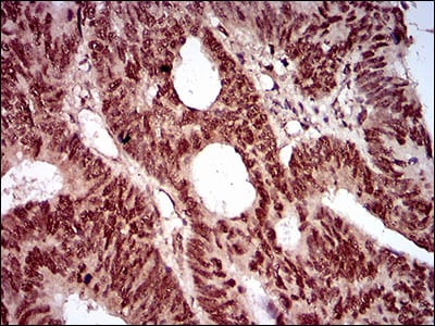
Figure 7: Immunohistochemical analysis of paraffin-embedded rectum cancer tissues using NBN mouse mAb with DAB staining.
Elisa

Black line: Control Antigen (100 ng); Purple line: Antigen(10ng); Blue line: Antigen (50 ng); Red line: Antigen (100 ng);
For Research Use Only. Not for use in diagnostic procedures.