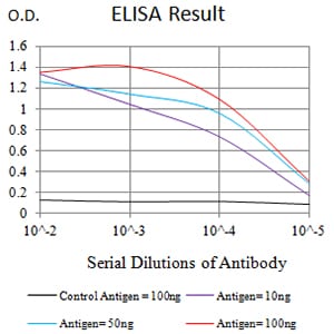
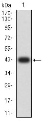
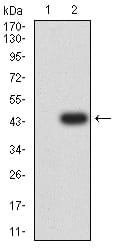
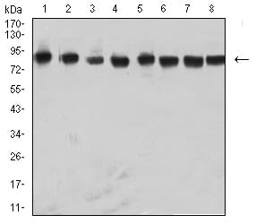
NAGR1 Primary Antibody
This gene belongs to the subfamily of ubiquitously expressed heterogeneous nuclear ribonucleoproteins (hnRNPs). The hnRNPs are RNA binding proteins and they complex with heterogeneous nuclear RNA (hnRNA). These proteins are associated with pre-mRNAs in the nucleus and appear to influence pre-mRNA processing and other aspects of mRNA metabolism and transport. While all of the hnRNPs are present in the nucleus, some seem to shuttle between the nucleus and the cytoplasm. The hnRNP proteins have distinct nucleic acid binding properties. The protein encoded by this gene has three repeats of quasi-RRM domains that bind to RNAs. This protein also constitutes a monomer of the N-acetylglucosamine-specific receptor which is postulated to trigger selective recycling of immature GlcNAc-bearing thyroglobulin molecules. Alternative splicing results in multiple transcript variants.
2.World J Gastroenterol. 2015 Feb 14;21(6):1784-93.
![Figure 1:Black line: Control Antigen (100 ng);Purple line: Antigen (10ng); Blue line: Antigen (50 ng); Red line:Antigen (100 ng)](https://cdn.shopify.com/s/files/1/0604/2661/7019/products/31503ea.jpg?v=1666703817)
![Figure 2:Western blot analysis using NAGR1 mAb against human NAGR1 (AA: 17-161) recombinant protein. (Expected MW is 42.2 kDa)](https://cdn.shopify.com/s/files/1/0604/2661/7019/products/31503rwb.jpg?v=1666703817)
![Figure 3:Western blot analysis using NAGR1 mAb against HEK293 (1) and NAGR1 (AA: 17-161)-hIgGFc transfected HEK293 (2) cell lysate.](https://cdn.shopify.com/s/files/1/0604/2661/7019/products/31503twb.jpg?v=1666703817)
![Figure 4:Western blot analysis using NAGR1 mouse mAb against Raji (1), Hela (2), NIH/3T3 (3), A431 (4), A549 (5), HepG2 (6), PC-12 (7), and U251 (8) cell lysate.](https://cdn.shopify.com/s/files/1/0604/2661/7019/products/31503lwb.jpg?v=1666703817)
![Figure 5:Immunofluorescence analysis of Hela cells using NAGR1 mouse mAb (green). Blue: DRAQ5 fluorescent DNA dye.](https://cdn.shopify.com/s/files/1/0604/2661/7019/products/31503icc.jpg?v=1666703817)
![Figure 6:Immunofluorescence analysis of Hela cells using NAGR1 mouse mAb (green). Blue: DRAQ5 fluorescent DNA dye. Red: Actin filaments have been labeled with Alexa Fluor- 555 phalloidin. Secondary antibody from Fisher (Cat#: 35503)](https://cdn.shopify.com/s/files/1/0604/2661/7019/products/31503icc-2.jpg?v=1666703817)
![Figure 7:Flow cytometric analysis of HL-60 cells using NAGR1 mouse mAb (green) and negative control (red).](https://cdn.shopify.com/s/files/1/0604/2661/7019/products/31503fcm.jpg?v=1666703817)
![Figure 8:Immunohistochemical analysis of paraffin-embedded esophageal cancer tissues using NAGR1 mouse mAb with DAB staining.](https://cdn.shopify.com/s/files/1/0604/2661/7019/products/31503ihc-1.jpg?v=1666703817)
![Figure 9:Immunohistochemical analysis of paraffin-embedded rectum cancer tissues using NAGR1 mouse mAb with DAB staining.](https://cdn.shopify.com/s/files/1/0604/2661/7019/products/31503ihc-2.jpg?v=1666703817)