

MUC2 Primary Antibody
Item Information
Catalog #
Size
Price
Description
This gene encodes a member of the mucin protein family. Mucins are high molecular weight glycoproteins produced by many epithelial tissues. The protein encoded by this gene is secreted and forms an insoluble mucous barrier that protects the gut lumen. The protein polymerizes into a gel of which 80% is composed of oligosaccharide side chains by weight. The protein features a central domain containing tandem repeats rich in threonine and proline that varies between 50 and 115 copies in different individuals. Downregulation of this gene has been observed in patients with Crohn disease and ulcerative colitis.
Product Overview
Entrez GenelD
4583
Aliases
MLP; SMUC; MUC-2
Clone#
4A5G8
Host / Isotype
Mouse / Mouse IgG1
Immunogen
Purified recombinant fragment of human MUC2 (AA: 4373-4557) expressed in E. Coli.
Formulation
Purified antibody in PBS with 0.05% sodium azide
Storage
Store at 4°C short term. Aliquot and store at -20°C long term. Avoid freeze/thaw cycles.
Product Applications
WB (Western Blot)
1/500 - 1/2000
FCM (Flow Cytometry)
1/200 - 1/400
ELISA
1/10000
References
1.Hum Pathol. 2014 Mar;45(3):540-8. 2.PLoS One. 2013 Dec 6;8(12):e79769.
Product Image
Elisa
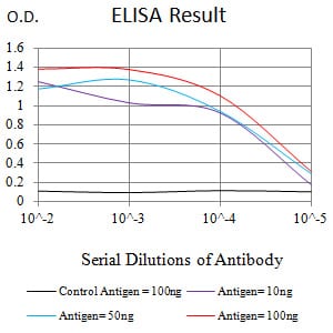
Figure 1:Black line: Control Antigen (100 ng);Purple line: Antigen (10ng); Blue line: Antigen (50 ng); Red line:Antigen (100 ng)
Western Blot
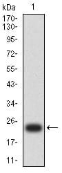
Figure 2:Western blot analysis using MUC2 mAb against human MUC2 (AA: 4373-4557) recombinant protein. (Expected MW is 23.8 kDa)
Western Blot
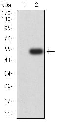
Figure 3:Western blot analysis using MUC2 mAb against HEK293 (1) and MUC2 (AA: 4373-4557)-hIgGFc transfected HEK293 (2) cell lysate.
Flow cytometric
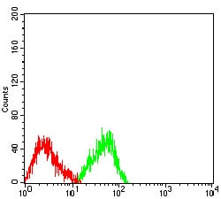
Figure 4:Flow cytometric analysis of SW480 cells using MUC2 mouse mAb (green) and negative control (red).
For Research Use Only. Not for use in diagnostic procedures.