

MUC13 Primary Antibody
Item Information
Catalog #
Size
Price
Description
Epithelial mucins, such as MUC13, are a family of secreted and cell surface glycoproteins expressed by ductal and glandular epithelial tissues (Williams et al., 2001 [PubMed 11278439]).
Product Overview
Entrez GenelD
56667
Aliases
DRCC1; MUC-13
Clone#
8B2C4
Host / Isotype
Mouse / Mouse IgG1
Species Reactivity
Human, Mouse
Immunogen
Purified recombinant fragment of human MUC13 (AA: extra 19-238) expressed in E. Coli.
Formulation
Purified antibody in PBS with 0.05% sodium azide
Storage
Store at 4°C short term. Aliquot and store at -20°C long term. Avoid freeze/thaw cycles.
Product Applications
WB (Western Blot)
1/500 - 1/2000
IHC_P(Immunohistochemistry)
1/200 - 1/1000
FCM (Flow Cytometry)
1/200 - 1/400
ELISA
1/10000
References
Micron. 2020 Mar;130:102822.
Int J Cancer. 2017 May 15;140(10):2351-2363.
Int J Cancer. 2017 May 15;140(10):2351-2363.
Product Image
Elisa

Figure 1:Black line: Control Antigen (100 ng);Purple line: Antigen (10ng); Blue line: Antigen (50 ng); Red line:Antigen (100 ng)
Western Blot
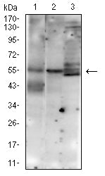
Figure 2:Western blot analysis using MUC13 mouse mAb against HT-19 (1), SW620 (2), and NIH/3T3 (3) cell lysate.
Immunofluorescence analysis

Figure 3:Flow cytometric analysis of LOVO cells using MUC13 mouse mAb (green) and negative control (red).
Immunohistochemical analysis

Figure 4:Immunohistochemical analysis of paraffin-embedded Colon cancer tissues using MUC13 mouse mAb with DAB staining.
Immunohistochemical analysis

Figure 5:Immunohistochemical analysis of paraffin-embedded rectal cancer tissues using MUC13 mouse mAb with DAB staining.
Immunohistochemical analysis

Figure 6:Immunohistochemical analysis of paraffin-embedded Rabbit small intestine tissues using MUC13 mouse mAb with DAB staining.
For Research Use Only. Not for use in diagnostic procedures.