

Mouse Monoclonal Antibody to MUC12
Item Information
Catalog #
Size
Price
Description
This gene encodes an integral membrane glycoprotein that is a member of the mucin family. Mucins are O-glycosylated proteins that play an essential role in forming protective mucous barriers on epithelial surfaces and have been implicated in epithelial renewal and differentiation. These glycoproteins also play a role in intracellular signaling. This protein is expressed on the apical membrane surface of epithelial cells that line the mucosal surfaces of many different tissues including the colon, pancreas, prostate, and uterus. The expression of this gene is downregulated in colorectal cancer tissue.
Product Overview
Entrez GenelD
10071
Aliases
MUC11; MUC-11;
Clone#
4A3D3
Host / Isotype
Mouse / IgG1
Immunogen
Purified recombinant fragment of human MUC12 (AA: extra 371-592) expressed in HEK293-6e cells supernatant.
Formulation
Purified antibody in PBS with 0.05% sodium azide
Storage
Store at 4°C short term. Aliquot and store at -20°C long term. Avoid freeze/thaw cycles.
Product Applications
WB (Western Blot)
1/500 - 1/2000
IHC_P(Immunohistochemistry)
1/200 - 1/1000
FCM (Flow Cytometry)
1/200 - 1/400
ELISA
1/10000
References
1.Mol Biochem Parasitol. 2019 Jan;227:19-24 2.Int J Cancer. 2010 Nov 15;127(10):2292-9.
Product Image
Elisa

Figure 1:Black line: Control Antigen (100 ng);Purple line: Antigen (10ng); Blue line: Antigen (50 ng); Red line:Antigen (100 ng)
Western Blot

Figure 2:Western blot analysis using MUC12 mAb against human MUC12 (AA: extra 371-592) recombinant protein. (Expected MW is 53.3 kDa)
Flow cytometric analysis
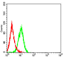
Figure 3:Flow cytometric analysis of Jurkat cells using MUC12 mouse mAb (green) and negative control (red).
Immunohistochemical analysis

Figure 4:Immunohistochemical analysis of paraffin-embedded stomach cancer tissues using MUC12 mouse mAb with DAB staining.
Immunohistochemical analysis

Figure 5:Immunohistochemical analysis of paraffin-embedded rectum cancer tissues using MUC12 mouse mAb with DAB staining.
For Research Use Only. Not for use in diagnostic procedures.