

MTDH Primary Antibody
Item Information
Catalog #
Size
Price
Description
MTDH (Metadherin) is a Protein Coding gene. Diseases associated with MTDH include Tongue Carcinoma and Gallbladder Adenocarcinoma. Among its related pathways are Gastric Cancer Network 2 and Validated targets of C-MYC transcriptional activation. Gene Ontology (GO) annotations related to this gene include double-stranded RNA binding. An important paralog of this gene is MAMSTR.
Product Overview
Entrez GenelD
92140
Aliases
3D3; AEG1; AEG-1; LYRIC; LYRIC/3D3
Clone#
8A4B12
Host / Isotype
Mouse / Mouse IgG1
Species Reactivity
Human, Monkey
Immunogen
Purified recombinant fragment of human MTDH (AA: 200-450) expressed in mammalian.
Formulation
Purified antibody in PBS with 0.05% sodium azide
Storage
Store at 4°C short term. Aliquot and store at -20°C long term. Avoid freeze/thaw cycles.
Product Applications
WB (Western Blot)
1/500 - 1/2000
IHC_P(Immunohistochemistry)
1/200 - 1/1000
FCM (Flow Cytometry)
1/200 - 1/400
ELISA
1/10000
References
1,Cell Death Dis. 2020 Apr 6;11(4):221.
2,Cancer Res. 2021 Feb 15;81(4):1014-1025.
2,Cancer Res. 2021 Feb 15;81(4):1014-1025.
Product Image
Elisa
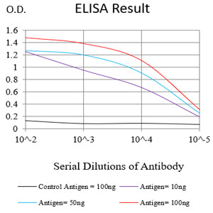
Figure 1:Black line: Control Antigen (100 ng);Purple line: Antigen (10ng); Blue line: Antigen (50 ng); Red line:Antigen (100 ng)
Western Blot

Figure 2:Western blot analysis using MTDH mAb against human MTDH (AA: 200-450) recombinant protein. (Expected MW is 57.8 kDa)
Western Blot

Figure 3:Western blot analysis using MTDH mouse mAb against MCF-7 (1), T47D (2), Jurkat(3), K562 (4), Hela (5),PC-3 (6), HepG2(7), A431(8) and COS7 (9) cell lysate.
Immunofluorescence analysis

Figure 4:Flow cytometric analysis of Hela cells using MTDH mouse mAb (green) and negative control (red).
Immunohistochemical analysis

Figure 5:Immunohistochemical analysis of paraffin-embedded bladder cancer tissues using MTDH mouse mAb with DAB staining.
Immunohistochemical analysis

Figure 6:Immunohistochemical analysis of paraffin-embedded prostate cancer tissues using MTDH mouse mAb with DAB staining.
For Research Use Only. Not for use in diagnostic procedures.