

MSN Primary Antibody
Item Information
Catalog #
Size
Price
Description
Moesin (for membrane-organizing extension spike protein) is a member of the ERM family which includes ezrin and radixin. ERM proteins appear to function as cross-linkers between plasma membranes and actin-based cytoskeletons. Moesin is localized to filopodia and other membranous protrusions that are important for cell-cell recognition and signaling and for cell movement.
Product Overview
Entrez GenelD
4478
Aliases
moesin
Clone#
2C12
Host / Isotype
Mouse / IgG1
Species Reactivity
Human, Monkey
Immunogen
Purified recombinant fragment of human MSN expressed in E. Coli.
Formulation
Purified antibody in PBS with 0.05% sodium azide
Storage
Store at 4°C short term. Aliquot and store at -20°C long term. Avoid freeze/thaw cycles.
Product Applications
WB (Western Blot)
1/500 - 1/2000
IHC_P(Immunohistochemistry)
1/200 - 1/1000
FCM (Flow Cytometry)
1/200 - 1/400
ELISA
1/10000
References
Int J Cancer. 2009 Apr 1;124(7):1614-21.
J Biol Chem. 2009 Jan 23;284(4):2419-34.
J Biol Chem. 2009 Jan 23;284(4):2419-34.
Product Image
Western Blot
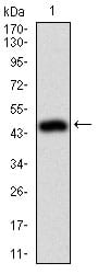
Figure 1: Western blot analysis using MSN mAb against human MSN (AA: 292-491) recombinant protein. (Expected MW is 49.2 kDa)
Western Blot

Figure 2: Western blot analysis using MSN mouse mAb against HeLa (1), A431 (2),Jurkat(3), HEK293(4), and COS7 (5) cell lysate.
Immunohistochemical analysis

Figure 3: Immunohistochemical analysis of paraffin-embedded colon tissues using MSN mouse mAb with DAB staining.
Flow cytometric

Figure 4: Flow cytometric analysis of Jurkat cells using MSN mouse mAb (green) and negative control (red).
Elisa
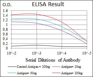
Black line: Control Antigen (100 ng); Purple line: Antigen(10ng); Blue line: Antigen (50 ng); Red line: Antigen (100 ng);
For Research Use Only. Not for use in diagnostic procedures.