

MSH6 Primary Antibody
Item Information
Catalog #
Size
Price
Description
This gene encodes a member of the DNA mismatch repair MutS family. In E. coli, the MutS protein helps in the recognition of mismatched nucleotides prior to their repair. A highly conserved region of approximately 150 aa, called the Walker-A adenine nucleotide binding motif, exists in MutS homologs. The encoded protein heterodimerizes with MSH2 to form a mismatch recognition complex that functions as a bidirectional molecular switch that exchanges ADP and ATP as DNA mismatches are bound and dissociated. Mutations in this gene may be associated with hereditary nonpolyposis colon cancer, colorectal cancer, and endometrial cancer. Transcripts variants encoding different isoforms have been described.
Product Overview
Entrez GenelD
2956
Aliases
GTBP; HSAP; p160; GTMBP; HNPCC5
Clone#
2E3A10
Host / Isotype
Mouse / Mouse IgG2b
Immunogen
Purified recombinant fragment of human MSH6 (AA: 374-540) expressed in E. Coli.
Formulation
Purified antibody in PBS with 0.05% sodium azide
Storage
Store at 4°C short term. Aliquot and store at -20°C long term. Avoid freeze/thaw cycles.
Product Applications
WB (Western Blot)
1/500 - 1/2000
IHC_P(Immunohistochemistry)
1/200-1/1000
FCM (Flow Cytometry)
1/200-1/400
ELISA
1/10000
References
1.Gene. 2019 Jul 1;704:103-112. 2.Biochem Biophys Res Commun. 2018 Feb 19;496(4):1040-1046.
Product Image
ELISA
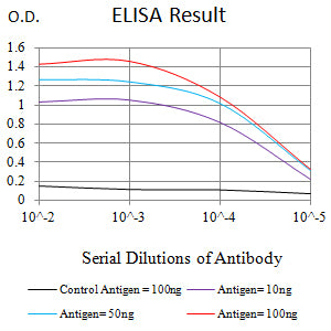
Figure 1: Black line: Control Antigen (100 ng);Purple line: Antigen (10ng); Blue line: Antigen (50 ng); Red line: Antigen (100 ng)
WESTERN BLOT
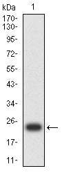
Figure 2: Western blot analysis using MSH6 mAb against human MSH6 (AA: 374-540) recombinant protein. (Expected MW is 22.6 kDa)
WESTERN BLOT
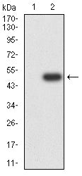
Figure 3: Western blot analysis using MSH6 mAb against HEK293-6e (1) and MSH6 (AA: 374-540)-hIgGFc transfected HEK293 (2) cell lysate.
WESTERN BLOT
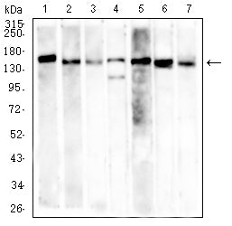
Figure 4: Western blot analysis using MSH6 mouse mAb against SH-SY5Y (1), K562 (2), Hela (3), PC-3 (4), HCT116 (5), HEK293 (6), and A549 (7) cell lysate.
FLOW CYTOMETRY
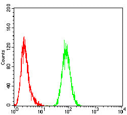
Figure 5: Flow cytometric analysis of Hela cells using MSH6 mouse mAb (green) and negative control (red).
IMMUNOHISTOCHEMISTRY
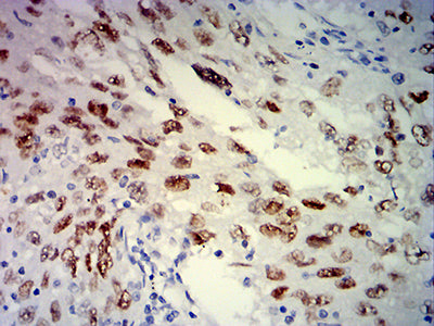
Figure 6: Immunohistochemical analysis of paraffin-embedded lung cancer tissues using MSH6 mouse mAb with DAB staining.
For Research Use Only. Not for use in diagnostic procedures.